Introduction
Nau mai, hoki mai, welcome back to your online study. This image header of a young girl's beautiful smile is the product of muscles, facial nerves and the brain working in harmony. This week, our travels continue through the human body, with a focus on muscle, bone, skin and hair (to name a few!). The endocrine system will also make a reappearance as we dive deep into the functions of the major glands in this system.
Tip
You will continue to be introduced to many new terms and definitions this week, so remember to add any unfamiliar terms to your glossary.
Feel free to include definitions in your own words or even draw/ source images to support the term. Explaining these concepts in your own words helps retention and understanding.
Hoake tatou, let’s go!
Welcome back to Patient Care 2. In this week’s session, we will explore the musculoskeletal and integumentary (skin) systems. We will look at their structure and function as well as some disorders.
Further learning on the disorders of the musculoskeletal and integumentary systems will be discussed in Anatomy and Physiology in another week.
The musculoskeletal system

The musculoskeletal system is made up of the skeletal and muscular systems working together to provide structure, support, and movement for the body. We will look at the major structures and functions of each system in turn.
The skeletal system
The skeletal system is made up of bones and joints. The main functions of this system are:
- Support: The skeleton provides support and structure to the body. Without bones, the human body would be like jelly!
- Movement: Bones provide attachment points for ligaments and tendons. Muscles are attached to bones via tendons. The bones act as rigid levers for movement to occur.
- Protection: Bones have the hugely important role of protecting the vital organs (think of the ribs as the bodyguards to the heart and lungs).
- Blood cell production: Red and white blood cells are formed in the bone marrow.
- Storage: Bones store calcium, phosphorus and fat tissue. Both can be released for growth and development and when the body needs extra energy.
Bones
There are 206 bones in the adult body that range in shape and size, but most of them are paired. That is, they have a ‘friend' on the other side of the body. What is on the left is typically on the right. However, babies and children have more bones than the adult human skeleton because as we grow, the bones fuse together (for example, bones of the skull).
Bones are also considered a living tissue as they perform a process called ‘remodelling’, which is the process of new bone forming and breaking down old bone. Pretty cool, huh?
There are five different shapes of bone:
- Flat: The sternum, ribs and most of the bones of the skull are flat bones.
- Long: Most bones of the limbs, including those of the fingers and toes, are long bones.
- Short: The bones of the wrist and ankle are short bones.
- Irregular: The bones of the spine, pelvis, and some bones of the skull are irregular bones.
- Sesamoid: Bones that are embedded in tendons. They hold the tendon away from the joint and increase the leverage of the muscle. An example is the patella.
Check out examples of these five shapes and their locations in the body below.

Bone isn’t just a single hard substance. Click the labels or (+) symbols to discover what bone consists of:
A fibrous membrane that covers the outer surface of the bone, it contains blood vessels, nerves, and cells important for bone growth, repair, and nutrition.
The dense outer layer of bone that surrounds the medullary cavity and contains blood vessels. It provides strength and rigidity.
The hollow part of the bone that contains yellow bone marrow. It produces cartilage, fat and bone. It also aids in the storage of fats in cells.
Found inside the bone, it is less dense than compact bone. As the name suggests, it consists of a spongy bone and contains red bone marrow, where blood cells are produced. Spongy bone contributes to the overall strength and flexibility of the bone.
Cartilage covers the end of bones. It is a tough, fibrous connective tissue that stops the bones from rubbing together and acts like a cushion to absorb shock.

Bone development and growth
Bone development starts early. Bones develop at about six weeks in the foetus and continue to grow until the age of 25. Most bones originate as hyaline cartilage. This cartilage is gradually converted to bone through a process called ossification. Bone growth activity occurs at the ends of the bones at the epiphyseal plate, commonly known as the growth plate.
The skeleton is subdivided into two parts that function in supporting the body, protecting vital organs, and facilitating movement.
These are the axial skeleton and the appendicular skeleton.

Axial skeleton
This is made up of the following:
- Skull: Flat bones that house and protect the brain.
- Vertebral column: Irregular bones, comprised of vertebrae, provide support and protect the spinal cord.
- Ribs: Flat bones that protect vital organs in the chest, such as the heart and lungs.
- Sternum: This may be referred to as the breastbone. It is a flat bone in the centre of the chest that connects to the ribs.
Appendicular skeleton
This is made up of the following:
- Upper limbs: Bones of the arms, including the humerus, radius, ulna, and hand bones (carpals, metacarpals, and phalanges). These are long and short bones.
- Lower limbs: Bones of the legs, including the femur, tibia, fibula, and foot bones (tarsals, metatarsals, and phalanges). These are long and short bones.
- Pectoral girdle: Includes the scapula (shoulder blade) and clavicle (collarbone) and connects the arms to the axial skeleton. These are flat bones.
- Pelvic girdle: The hip bones. It supports the lower limbs and protects internal organs. These are irregular bones.
Watch: Functions Of Bones In The Body […] (1:29 minutes)
Before we move on to the next component of the musculoskeletal system, let’s recap the types and functions of bones. Mātakitaki mai, watch for an explanation of the main functions and then complete the activity to check your understanding.
Joints
A joint is a point where bones meet and allow for movement:
- between two bones
- between bone and cartilage
- between bone and teeth.
The two main functions of joints are:
- Hold the skeleton together.
- Give the skeleton mobility.
Ligaments, tendons, and cartilage are all integral components of the joint system, working together to support and facilitate movement within the skeletal system.
Expand the labels below for a description of each of these.
These are strong, fibrous tissues that connect bones to bones. They provide stability to joints and prevent excessive movement.
These are fibrous tissues that connect muscles to bones. Tendons transmit the force generated by muscles to bones, allowing for movement.
Found at the ends of bones in joints, cartilage acts as a cushion, reducing friction and preventing wear and tear during movement. It also contributes to the flexibility and smooth functioning of joints.
Ligament vs. tendon
| Here, you can see the differences between a ligament (bone connecting bone) and a tendon (muscle connecting bone). | 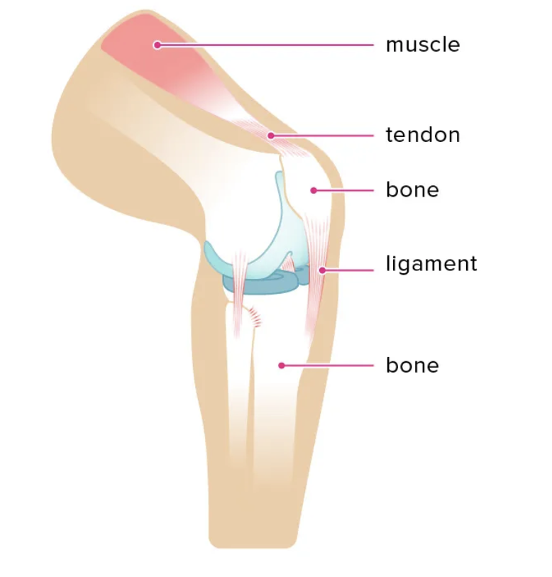 |
Types of joints
Joints are grouped according to their type of motion, and most are even named accordingly (making them easier to remember!).
Ball and socket joint
The end of one bone has a 'ball-like' end, and the other bone has a 'socket' that the ball sits in. They have the greatest range of motion in the human body.
Examples - hip and shoulder joints.
Hinge joint
Permits movement in only one plane.
Example - elbow.
Condyloid joint
Shallow ball and socket joints that can do everything a ball and socket joint can do except rotate.
Example - base of the index finger, elbow and wrist joints.
Pivot joint
A pivot is a pure 'rotating' joint.
Example - neck, between C1 and C2 vertebrae.
Plane or gliding joint
It occurs when short bones glide along each other in all directions (but not far). Think of it like sliding two coins together between your fingers.
Examples - wrist and ankle.
Saddle joint
A saddle is when the end of one bone is like a concave 'saddle' that the other bone sits over like two legs. It allows a substantial range of movements. The concave and convex surfaces fit together.
Example - base of thumb and wrist.
Check out this image that illustrates the movement of these joints and their locations in the body.

The muscular system
This system is made up of tissues that have the unique ability to contract and produce movement.
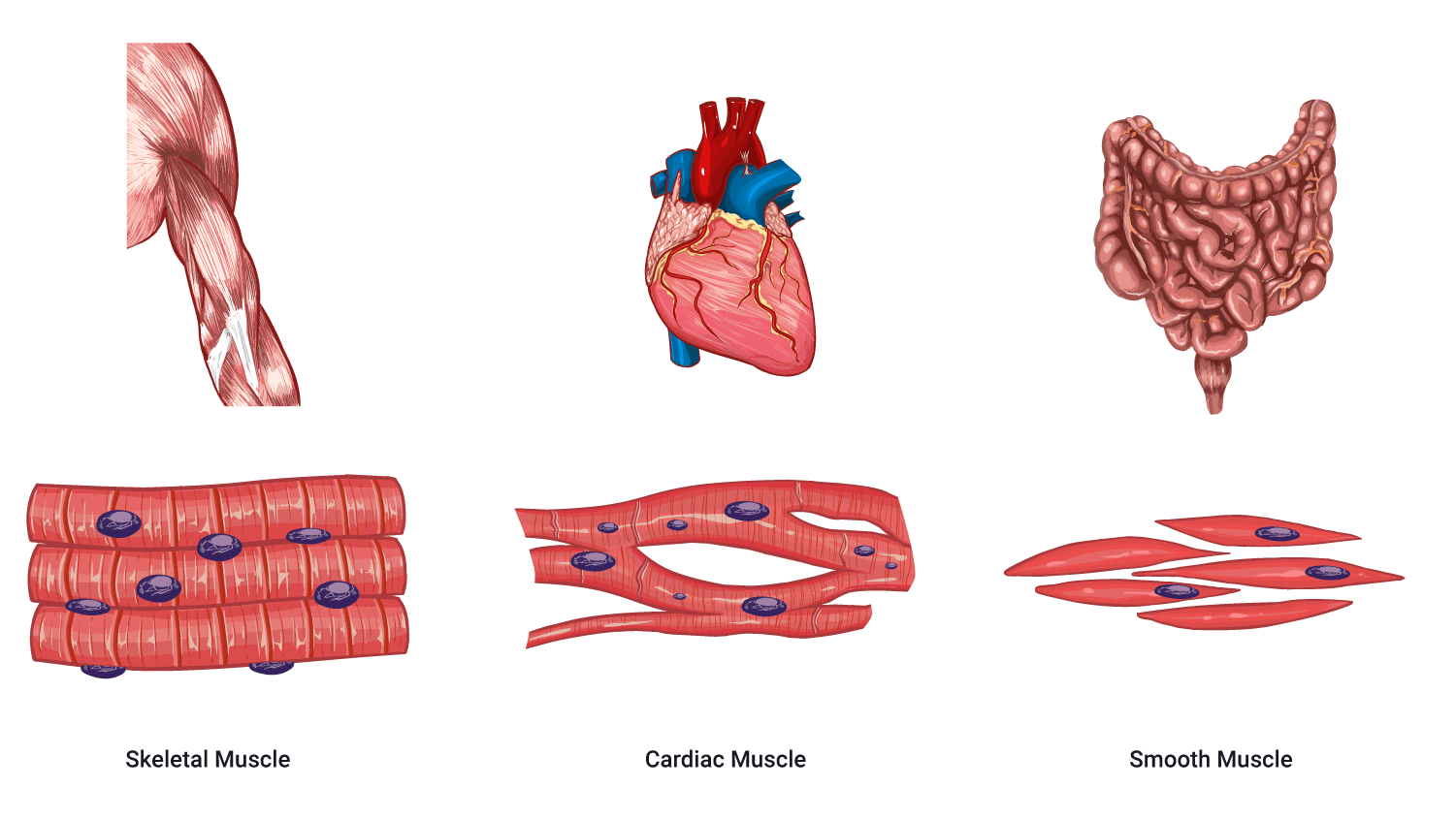
There are three types of muscle tissues, these are:
- Skeletal muscle tissue
- Smooth muscle tissue
- Cardiac muscle tissue
The following image shows the differences between the three types.
Characteristics of muscle tissue
Although the names are fairly self-explanatory, let’s identify some important points about each muscle type.
Skeletal muscle tissue
- These are the muscles attached to bones by tendons, hence the name ‘skeletal.’
- Skeletal muscles are bundles of contractile fibres that are organised in a regular pattern so that under a microscope, they appear as stripes (which is why they are also called striped or striated muscles).
- Skeletal muscles vary in their speeds of contraction. Most of these muscles are under voluntary control, which means that it is mostly consciously controlled by the somatic nervous system.
- Their contraction allows for conscious movements, such as walking, running, and lifting objects.
Fun fact: Skeletal muscle makes up approximately 40% of body weight.
Smooth muscle tissue
- Named for its smooth appearance under a microscope.
- Found in the walls of internal organs, such as the digestive tract, blood vessels, and respiratory passages.
- Smooth muscles operate involuntarily. They are regulated by the autonomic division of the nervous system, which means that you can’t consciously control it.
- They are responsible for various internal processes, including peristalsis in the digestive system.
Cardiac muscle tissue
- Exclusive to the heart, cardiac muscles contract rhythmically to pump blood throughout the circulatory system.
- Like skeletal muscle, cardiac muscle is striated - it has a regular pattern of fibres that appear as stripes under a microscope.
- Like smooth muscles, cardiac muscles work involuntarily, ensuring the continuous circulation of blood to meet the body’s needs. This is lucky – imagine having to think of contracting your heart to pump blood around your body!
Watch: How your muscular system works – Emma Bryce (4:44 minutes)
Did you know that almost every motion the human body makes is governed by the muscular system? Muscles are responsible for allowing humans to walk, blink, hold a cup of coffee and even give birth!
Mātakitaki mai, watch this video and take note of how the muscular system works and what parts of the nervous system are involved in its functioning. Complete the activity following this video to connect your knowledge of these two systems and broaden your learning.
The musculoskeletal system is a partnership between the skeletal and muscular systems. This partnership enables the body to move, support itself and carry out a variety of body functions.
Musculoskeletal disorders and treatment
We will look more widely at disorders and treatment in Anatomy and Physiology, but for now, let’s review what you already know about two very common muscular skeletal injuries - strains and sprains.
Journal post
Musculoskeletal disorder and treatment: Strain and Sprain
What do you already know about strains and sprains?
- Download and complete this worksheet using your prior knowledge: Week 30 - MS Disorder and Treatment: Strains and Sprains.
- Note: If you need a hand answering questions, speak to a classmate or conduct research.
- Upload the completed worksheet to a new journal post titled ‘Musculoskeletal Disorder and Treatment: Strains and Sprains’.
- Publish the post to ‘All course users’ as your tutor will check your work.
- Save the permalink to your Index of Journal Posts.
The integumentary system

The integumentary system is the set of organs that form a layer to keep our insides in, and everything else out.
It’s made up of the following structures:
- Skin
- Hair
- Nails
- Sebaceous glands
- Sweat glands
- Blood vessels
- Nerve endings
Skin
The skin is the largest organ of the body and serves as a protective barrier and temperature regulator. Also known as the cutaneous membrane, it is composed of three layers:
- the epidermis (the outer layer).
- the dermis (the inner layer).
- the hypodermis (which is sometimes referred to as the subcutaneous layer).
Hair
Hair are structures composed of keratin that grow from hair follicles in the skin. Hair provides insulation and protection.
Nails
Nails are hard structures made up of keratinised cells that cover the tips of fingers and toes. They provide protection and support.
Sebaceous glands
These are oil-producing glands located in the skin that secrete sebum. They help to lubricate and waterproof the skin and hair, keep the skin supple and make the skin inhospitable to foreign organisms.
Sweat glands
Sweat glands help to regulate body temperature through evaporative cooling and eliminating waste products.
There are two types of sweat glands that produce sweat:
- Eccrine glands: open directly to the skin surface.
- Apocrine glands: open into the hair follicle, leading to the skin surface.
Blood vessels
Although blood vessels are part of the cardiovascular system, they play a vital role in the integumentary system. The network of blood vessels in the skin supplies nutrients and oxygen while regulating temperature.
Nerve endings
Nerve endings are parts of the nervous system that have an important role in the integumentary system. The nerve endings in the skin are crucial for sensing the external environment and relaying information about various stimuli to the brain for processing and response.
Fun fact: These nerve endings and receptors are finely tuned to let humans feel the world around them, be it a slight breeze on your skin or the feeling of touching a hot pan. Weirdly, though, we don’t have any receptors for wetness. This is why when you sit on a wet bench, you can’t tell if it’s wet or just cold.
The skin’s anatomy
Take a moment to identify these structures and become familiar with the organisation of the integumentary system.

Integumentary system functions
Use your prior knowledge and the content you’ve just read to complete this quick activity.
How did you go? Do you know how each of the functions of the integumentary system is achieved? If not, it would be a good idea to carry out your own search for this information.
For example, for the function of ‘Protection’, ask yourself, “How does the integumentary system function as protection?” Your answer would be, “The skin acts as a covering and physical barrier, protecting the body from external threats such as pathogens, chemicals, and physical injuries.”
Integumentary disorders and treatment
As this is a body system we will look at more widely in our Anatomy and Physiology learning, we will only briefly touch on disorders and treatments now, starting with a fun activity.
Acne and dermatitis activity
Two integumentary disorders that you will be familiar with from your previous learning are dermatitis and acne. Both skin conditions are common and can flare, go away for a while and return.
The purpose of this activity is to get you thinking about the role the integumentary system plays in these two very common disorders. Don’t worry if you don’t get all the answers correct. The objective is to confirm what you already know and to encourage you to consciously take note of anything that you’re not confident about.
Karawhiua, give the activity a go!
Burns
You may remember learning about the different causes and types of burns in your previous learning in this programme. This next learning activity will help you to check what you know. Feel free to carry out some research to help refresh your memory.
Self-directed learning activities
How confident do you feel about the anatomy and basic functions of the musculoskeletal and integumentary body systems? Complete these three activities to recap this week’s content and build on your understanding.
Activity 1 - Muscular system
Although we touched on the types of muscles in the body, we didn’t delve into the names of the main muscles in the body.
Use your current knowledge to have a go naming the muscles in this illustration.
No worries if you didn’t get them all correct! Keep retrying the activity until you have nailed them all, and then download and save the labelled illustration as a resource: Week 30 – Patient Care 2 – Muscular System.
Activity 2 - Integumentary system: Disorders
The following two videos summarise your learning of dermatitis and acne. We encourage you to write notes as you watch. You might like to list examples, paraphrase the key information or even draw diagrams or mindmaps to help you memorise and connect this information to your existing knowledge.
Watch: Dermatitis vs Eczema, are they the same? (3:55 minutes)
Watch: Acne: Understanding the Types of Acne and Treatment Options (5:43 minutes)
Click this link to visit YouTube to watch the video.
Activity 2 - Level up (Optional)
What is one new piece of information you can recall from each video?
Answer this question (either in your head or in your notes).
By asking yourself questions like this, you create an awareness and understanding of your learning process. If you don’t feel like you learned anything, consider trying a different note-taking technique.
Activity 3 - Integumentary system: Burns
Our friend Abraham, The Pharmacist, joins us again as he shares guidance on how to treat burns and scalds and, most importantly, when to seek medical advice.
Watch the video and then answer the journal post question that follows.
Watch: Burns | How To Treat Burns (4:30 minutes)
After you’ve watched the video:
- Create a new journal post titled ‘SDL - Integumentary system: Burns’
- Answer this question: When would you advise a patient with a burn to seek medical attention?
- Publish the post to ‘All course users’.
- Save the permalink to your Index of Journal Posts.
Whakamihi, congratulations, you’ve completed Week 30 of Patient Care 2. Ko koe a runga, you’re awesome.

Welcome to Week 30. This session will delve further into the endocrine system.
Me ruku ki te kaupapa, let’s dive into the topic!
Endocrine system
In Week 28 of Patient Care 2, you were introduced to the endocrine system. You watched a video and answered some questions about the structure and function of this system.
Endocrine system review
To refresh your learning before we move on, we encourage you to revisit the following:
- YouTube video: GCSE Biology – Endocrine System & Hormones #59.
- Your journal post, titled ‘The Endocrine System’.
- Your SDL activity in your journal post, titled ‘SDL - Endocrine Disorder’.
Complete the following activity to recap and revise some key points about the endocrine system:
Endocrine glands

You will remember this diagram from the Week 28 journal post activity. This week, we will take a closer look at these (mostly tiny) endocrine glands and the huge importance that they have on happiness and well-being through the hormones that they produce.
Hypothalamus
The hypothalamus is a pea-sized region of the brain located behind the eyes and is a vital component of both the nervous system and the endocrine system. The hypothalamus maintains homeostasis by responding to chemical nerve signals from the central and peripheral nervous system. It achieves this by directly influencing the autonomic nervous system and managing the release of hormones.
Hormones
The hypothalamus makes the following hormones:
- Oxytocin
- Antidiuretic hormone
- Releasing hormones:
- Thyrotropin-Releasing Hormone (TRH)
- Corticotropin-Releasing Hormone (CRH)
- Gonadotropin-Releasing Hormone (GnRH)
- Growth Hormone-Releasing Hormone (GHRH)
- Prolactin-Releasing Hormone (PRH)
- Inhibiting hormones:
- Prolactin-Inhibiting Hormone (PIH) or Dopamine
- Growth Hormone-Inhibiting Hormone (GHIH) or Somatostatin
Pituitary gland
The hypothalamus closely interacts with the pituitary gland. Located at the base of the brain, a short stalk of tissue connects the pituitary gland to the hypothalamus.
Although the pituitary gland is only about the size of a baked bean, it is often called the master gland as it controls so much and has an impact on almost every part of your life. It produces (or regulates) the production of oestrogen, testosterone, cortisol and adrenaline (just to name a few).

It is divided into two parts: the anterior (front) pituitary and the posterior (behind) pituitary.
Posterior pituitary (Neurohypophysis)
The posterior pituitary is composed of nervous tissue and contains the terminations (ends) of nerve fibres that originate in the hypothalamus.
Hormones
The posterior pituitary stores the oxytocin and antidiuretic hormone (ADH) (sometimes called ‘vasopressin’) made by the hypothalamus. When the nerve fibres are stimulated, these hormones are released directly into the bloodstream.
Anterior pituitary (Adenohypophysis)
The anterior pituitary is made of different cell types that produce and release various hormones. The hypothalamus tightly regulates the production and release of these hormones, which travel through blood vessels to the anterior pituitary, where they stimulate or inhibit the release of specific hormones into the bloodstream.
Hormones
These hormones are:
- Growth Hormone (GH)
- Thyroid-Stimulating Hormone (TSH)
- Adrenocorticotropic Hormone (ACTH)
- Luteinizing Hormone (LH)
- Follicle-Stimulating Hormone (FSH)
- Prolactin (PRL)
Want to know what the pituitary gland has to do with the Guinness World Records? Click the (+) symbol to read a radical example of how a pituitary gland condition impacted the tallest human who ever lived.
Robert Wadlow was the tallest person ever to live, reaching 8 ft 11.1 inches (2.72m) when he sadly passed away at 22. That’s taller than a grizzly bear standing on its hind legs! Robert’s towering height was caused by a pituitary condition called hypertrophy, which means his gland was bigger than expected and caused a continuous overproduction of GH. This meant that he never stopped growing.
If you’re interested in learning more about Robert and why he is likely to be the tallest man ever, read this article from Guinness World Records: Why Robert Wadlow will be the tallest person, ever, forever.
Journal post
Hypothalamus and pituitary hormones
It’s time to use your pūkenga rangahau (research skills) to find out the actions of the following hormones.
- Create a new journal post titled ‘Hypothalamus and Pituitary Hormones’.
- For each hormone listed below, research its action and share your findings. You can format your findings as you wish.
- Publish your post to ‘All course users’.
- Save the permalink to your Index of Journal Posts. Your tutor may review your mahi, so remember to check back on your post in the coming weeks for feedback.
Hormones
- Hormones secreted by the hypothalamus:
- Prolactin-Inhibiting Hormone (PIH)
- Growth Hormone-Inhibiting Hormone (GHIH)
- Hormones secreted by the anterior pituitary:
- Growth hormone (GH)
- Thyroid-stimulating hormone (TSH)
- Adrenocorticotropic hormone (ACTH)
- Luteinizing Hormone (LH)
- Follicle-Stimulating Hormone (FSH)
- Prolactin (PRL)
- Hormones secreted by the posterior pituitary:
- Oxytocin
- Antidiuretic hormone (ADH)
Pineal gland

The pineal gland is a small, cone-shaped located deep within the brain, between the two hemispheres.
Hormones
The pineal gland secretes the hormone melatonin in response to low light levels. Melatonin regulates the sleep-wake cycle (circadian rhythm).
Thyroid gland

The thyroid gland is in the lower part of the neck below the Adam's apple, wrapped around the trachea. It consists of two lobes connected by a narrow band of tissue called the isthmus.
Hormones
The thyroid gland is composed of follicular cells that produce and store the thyroid hormones thyroxine (T4) and triiodothyronine (T3). These hormones are produced and secreted in response to thyroid-stimulating hormone (TSH) from the anterior pituitary gland and are involved in regulating metabolism, growth, development, and body temperature.
The thyroid gland also produces the hormone calcitonin in response to high levels of calcium in the blood. Calcitonin acts to lower blood calcium levels by blocking the activity of cells that break down and release calcium into the blood, inhibit calcium absorption into the blood from the digestive and urinary systems and promote calcium absorption from the blood into the bones.
Watch: Thyroid gland – What’s the function of the thyroid? (1:54 minutes)
This video illustrates the function of the thyroid gland, its relationship to the pituitary and the role of thyroxine (T4) and triiodothyronine (T3). We encourage you to make your own notes about the structure and function of the thyroid gland to help you cement your learning.
Parathyroid glands

These are small, pea-sized glands located on the back surface of the thyroid gland. There are typically four parathyroid glands, but the number can vary.
Hormones
The parathyroid glands are composed of cells that produce parathyroid hormone (PTH), which regulates blood calcium levels. Low blood calcium levels cause the production and secretion of PTH. In contrast, elevated blood calcium levels inhibit the secretion of PTH and trigger the thyroid to produce calcitonin.
Thymus gland

The thymus gland is part of the endocrine and lymphatic body systems. It is in the upper part of the chest, behind the sternum (breastbone) and in front of the trachea. Interestingly, it is most prominent during childhood and begins to decrease in size and activity during adolescence.
The thymus is composed of two lobes and is subdivided into lobules, each containing a cortex and a medulla.
Hormones
The thymus produces the hormone thymosin and other hormones that are essential in the development and maturation of T lymphocytes (T cells), a type of white blood cell involved in the immune response.
Adrenal glands

The adrenal glands are a pair of small, triangular-shaped glands located on top of each kidney. Each adrenal gland is divided into two distinct regions:
- Adrenal cortex
- Adrenal medulla
Adrenal cortex
- The outer layer of the adrenal gland.
- Produces steroid hormones, including cortisol, aldosterone, and androgens.
Adrenal medulla
- The inner core of the adrenal gland.
- Functions as part of the sympathetic nervous system.
- Produces catecholamines, such as adrenaline (epinephrine) and noradrenaline (norepinephrine).
Journal post
Adrenal hormones
Once more, it’s time to learn by doing your own rangahau (research) on the actions of adrenal hormones.
-
Create a new journal post titled ‘Adrenal Hormones’.
-
For each of the following adrenal hormones, research its action and share your findings. You can format your findings as you wish.
-
Publish your post to ‘All course users’.
-
Save the permalink to your Index of Journal Posts. Your tutor may review your mahi, so remember to check back on your post in the coming weeks for feedback.
Hormones
- Aldosterone
- Cortisol
- Adrenaline (epinephrine)
- Noradrenaline (norepinephrine)
Pancreas

You will recognise this illustration from Week 28 of Patient Care 2, where we noted that the pancreas plays a role in the digestive and endocrine systems. This week, we are focusing on the endocrine function of this organ.
Pancreatic islets
Throughout the pancreas, there are clusters of cells known as pancreatic islets, formerly known as the Islets of Langerhans. The islets contain different cell types, each producing specific hormones. We will focus on the alpha and beta cell types, the hormones they produce and how they work together to regulate blood sugar levels, contributing to the body's overall state of homeostasis.
Terminology
Before we go any further, it’s important to understand the terminology we will be using. There are three words that start with the letter ‘g’ and sound similar, so they are easy to confuse!
Click the ‘+’ icon for more information on each ‘G’.
- Glucose is a simple sugar, a fundamental source of energy for the body's cells.
- It is a primary fuel for various cellular processes, especially in the brain.
- Blood glucose levels are tightly regulated to ensure a steady and adequate supply of energy.
- Glycogen is a complex carbohydrate and is a storage form of glucose found in the liver and muscles.
- It serves as a reserve of glucose for when the body needs an additional supply of energy, helping to maintain blood glucose levels during fasting or between meals.
- Glucagon is a hormone produced by the alpha cells of the pancreas.
- It acts to increase blood glucose levels.
- It stimulates the breakdown of glycogen into glucose (glycogenolysis) in the liver, releasing glucose into the bloodstream.
- Additionally, it promotes gluconeogenesis, the production of glucose from non-carbohydrate sources.
Confused? Ensure that you understand the differences between each word before continuing. You may wish to add the terms to your glossary in your own words, create a table defining each term, draw your own diagrams, or do a Google search for illustrations or examples to add to your notes.
Alpha and beta cells
In the pancreas, alpha cells produce the hormone glucagon, which increases blood glucose levels by promoting the breakdown of glycogen (the stored form of glucose).
Conversely, beta cells produce insulin, a hormone that lowers blood glucose levels by facilitating glucose uptake into cells. Insulin is often described as the ‘key’ that unlocks the cell to allow glucose to enter. Without insulin the glucose in the bloodstream cannot enter the cell.
Watch: GSCE Biology – Control of Blood Glucose Concentration #56 (4:33 minutes)
Āta mātakitaki, watch this video carefully for an explanation of blood glucose control. Make sure to focus on the explanation near the end of the ‘negative feedback loop’ that ensures blood glucose levels remain within the normal range. You will examine and apply this explanation to the following activity.
Journal post
Blood Glucose Control

- Looking at this illustration and using what you have learnt from the video, create your own explanation of how the pancreas responds to the following situations:
- Low blood glucose.
- High blood glucose.
- In your explanation, make sure to:
- name the hormones released by the pancreas.
- explain how they act to return blood glucose levels to normal.
- Post your explanation to a new journal post titled ‘Blood Glucose Control’.
- Publish to ‘All course users’ so your tutor can review your mahi.
- Save the permalink to your Index of Journal Posts.
Ovaries and testes

Can you locate the ovaries and testes in this illustration?
As you will remember from our discussion on the reproductive system, the male gonads are the testes, and the female gonads are the ovaries. The gonads, as well as belonging to the reproductive system, also belong to the endocrine system as they secrete hormones.
These hormones prepare the body for reproduction and regulate various physiological processes, including the development of secondary sexual characteristics, menstrual cycles in females, and the production of sperm in males.
Ovaries
The primary hormones of the ovaries are oestrogen and progesterone.
Oestrogen
Oestrogen contributes to the development of secondary sexual characteristics during puberty, such as breast development and the widening of the hips and plays a central role in the regulation of the menstrual cycle, including the growth and development of the uterine lining (endometrium) during the menstrual cycle.
Progesterone
Progesterone works with oestrogen to prepare and maintain the uterine lining for possible implantation of a fertilised egg. If pregnancy occurs, progesterone helps maintain the uterine environment suitable for foetal development. It also prevents the uterine muscles from contracting, preventing premature labour.
Ovarian hormones also have effects beyond reproduction. They influence bone density, cardiovascular health, and the overall well-being of various tissues and organs in the female body.
Testes
The primary hormone produced by the testes is testosterone.
Testosterone
Testosterone is crucial for the development and maturation of sperm cells (spermatogenesis) in the testes. It is responsible for the development of secondary sexual characteristics during puberty, such as the deepening of the voice, facial and body hair growth, and increased muscle mass. Testosterone also supports the maintenance of male reproductive organs, including the testes and prostate.
Summary
In this session we have looked at the structure and function of the endocrine system. We have learnt that the endocrine system is a complex network of glands and organs that produce and release hormones, and act as chemical messengers that regulate various processes in the body.
These hormones travel through the bloodstream to target cells or organs, where they act to maintain homeostasis, regulate growth and development, control metabolism, and influence various other functions crucial for overall health and well-being. The endocrine system works in coordination with the nervous system to ensure proper communication and control of bodily functions.
Self-directed learning activity
In your SDL activity for this week, you will explore three common endocrine disorders of the thyroid gland and create your own learning material.
Watch: Thyroid problems – most common thyroid problems, symptoms and treatment (4:10 minutes)
This animation covers the symptoms and treatment of hyperthyroidism, hypothyroidism, and goitre. Take notes as you watch, then complete the following activity.
And just like that, you’ve completed Week 30. Galuega lelei, good job.