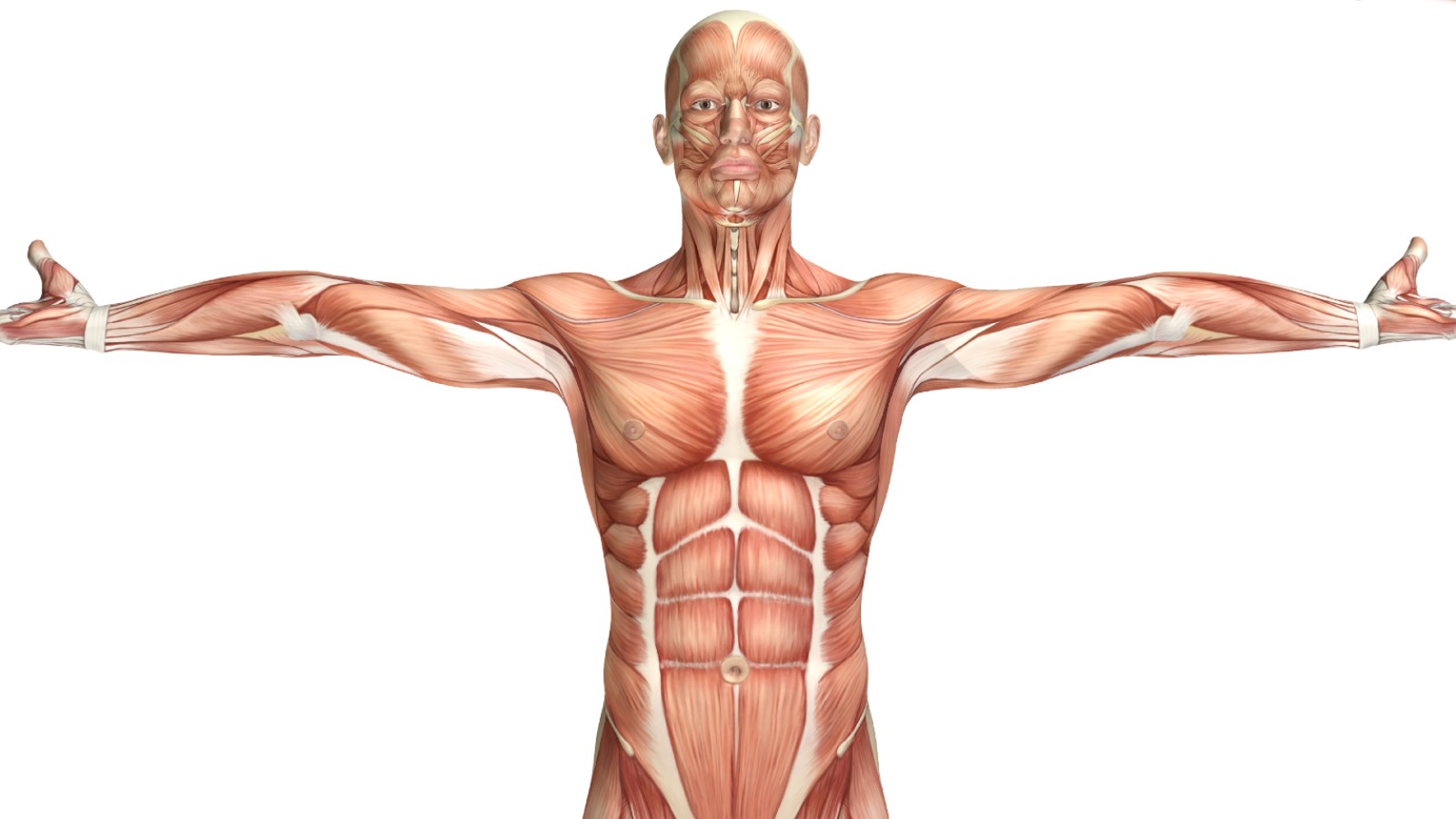
Having a general understanding of how the human body is constructed and how each part and system works will help you to understand how medical considerations should be accounted for, how injuries may be caused, and what sort of adaptations will result from regular exercising.
The human body consists of about 60% water. Along with water, other organic compounds such as lipids, proteins, carbohydrates and nucleic acids contribute to the majority of the human body composition.
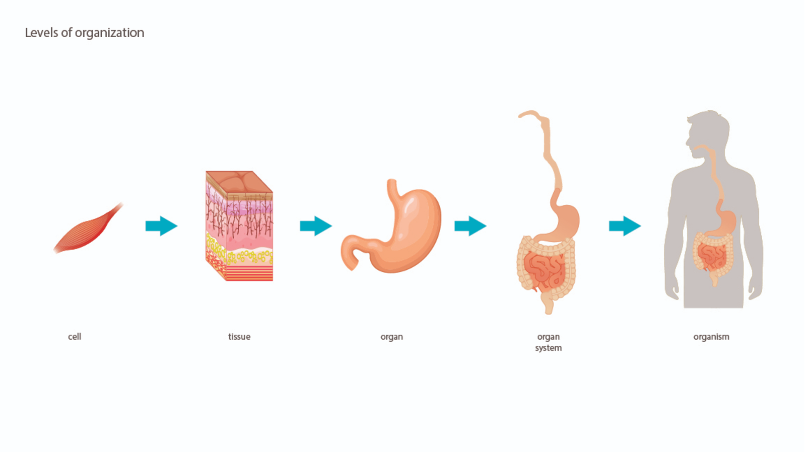
The structural levels of organisation of the human body are:
-
Cell.
-
Tissue.
-
Organ.
-
Organ system.
-
Organism.
The human body consists of trillions of cells, they are the basic living unit of the human body (likewise all organisms). Cells are capable of growth, metabolism, response to stimuli and reproduction (with some exceptions).
There many different types of cells in the body, however, they can be grouped into four basic classes. These classes, along with extracellular materials, form the fundamental tissues of the human body:
-
Epithelial Tissues - Cover the body's surface and line the internal organs, body cavities, and passageways. For example, the outer layers of the skin consists of epithelial tissue.
-
Muscle Tissues - Capable of contraction and form the body's musculature. For example, skeletal muscles like the bicep consists of muscle tissue.
-
Nerve Tissues - Conduct electrical impulses and make up the nervous system. For example, the spinal cord consists of nerve tissue.
-
Connective Tissues - Composed of widely spaced cells and large amounts of intercellular matrix. This type of tissue binds together various body structures. For example, blood and bone are a types of connective tissue.
An organ is a group of tissues which have a structural and functional purpose. The human body consists of many different organs, the exact number of which varies because the definition of what constitutes an organ is debated.
Examples of organs are:
-
Skin - The largest organ and outer layer in the human body. Among its many functions, it provides protection, sensation, heat regulation, evaporation control and water resistance.
-
Heart - An organ about the same size as the adult fist which pumps blood throughout the body.
-
Lungs - Pyramid-shaped organs within the chest which bring oxygen into the body through the breathing process and expel carbon dioxide out.
-
Liver - A large organ weighing approximately 1.3kg, it converts nutrients into substances the body is able to use and stores these substances until they are needed. It also functions as a blood detoxification and purification system.
-
Stomach - A muscular hollow organ that serves three main purposes: Temporary storage of food (2+ hours), mixing of the food items, and breaking down of the food by contraction and relaxation of its muscle layers.
-
Brain - The brain controls thoughts, memory, speech, movement and the function of organs within the body. It also interprets information from the outside world (via senses).
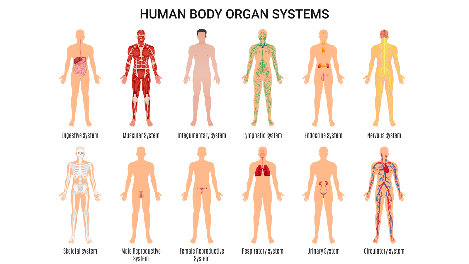
There are either eleven or twelve organ systems within the human body. This varying number depends on whether you combine the Immune system and the Lymphatic system together (as they work in conjunction with each other). For the purposes of this topic they have been separated). Each system contains several specific organs and works together to maintain a healthy body.
In alphabetical order they are:
Made up of the blood vessels, blood, and the heart it is responsible for distributing (or circulating) a range of nutrients, oxygen, hormones and waste products around the body, contributing to a healthy body by ensuring that all of the other organs, muscles, and tissues have the items that they need in order to survive and thrive and waste products are removed.
The heart is a muscular pumping organ located medially (towards the midline) within the thoracic cavity, intermediate (between) the lungs, deep (behind/farther from the surface) and slightly to the left of the breastbone (sternum). The heart consists of four chambers through which blood flows.
-
Right Atrium - Blood returning from the body enters the heart via this chamber, it then moves into the second chamber.
-
Right Ventricle - Blood is pumped through this chamber on its way to the lungs to become oxygenated. This process is known as Pulmonary Circulation.
-
Left Atrium - Oxygenated blood returning from the lungs passes back through this chamber.
-
Left Ventricle - This chamber pumps the blood to the Aorta which distributes it around the body. This process is known as Systemic Circulation.
There are four valves that work in conjunction with the chambers of the heart to ensure that blood is pumped around the heart in the correct direction, the are like gates that only open one way. These valves are:
-
Tricuspid Valve - As the Right Atrium fills up this valve opens to allow blood to flow into the Right Ventricle.
-
Pulmonary Valve - Connects the Right Ventricle and the Pulmonary Artery.
-
Mitral Valve - Located between the Left Atrium and Left Ventricle it regulates blood flow from the lungs into the Left Ventricle.
-
Aortic Valve - Allows blood to flow into the Aorta (the largest artery in the human body) and then onto the rest of the body.
The pumping action of the heart is a muscle contraction and relaxation process, the contraction action is known as Systole and the relaxation is known as Diastole. A series of electrical impulses deliver the signal for the heart to pump in a coordinated and normal rhythm.
The role of blood is to transport oxygen, cells, protein, water, nutrients and waste around the body so it can be utilised or cleansed by various organs. Blood is distributed around the body via arteries, veins and capillaries, which are known collectively as blood vessels. This distribution process is called circulation. It takes less than 60 seconds to circulate blood to all of the body's cells. The difference between blood vessels are:
-
Arteries - Carry oxygen rich blood away from the heart and circulate it around the body.
-
Veins - Bring the blood back to the heart so that it can be oxygenated again and continue the journey.
-
Capillaries - Tiny blood vessels which help to connect arteries to veins, transfer nutrients from the blood to various tissues and collect carbon dioxide waste.
The heart must also supply blood, oxygen, nutrients, and remove waste products within itself to sustain its cardiac cells (cardiomyocytes). This process is known as Coronary Circulation and it involves a network of arteries and veins located specifically within the heart muscle.
The video below provides a visual overview of all the process discussed above.
In relation to fitness, the circulatory/cardiovascular system delivers oxygenated blood to muscles during aerobic activities such as walking, jogging or cycling. Prior to exercise, the resting heartbeat rate is usually around 60-80 times per minute. As the intensity of exercise increases, this heart reacts and works harder to supply the blood faster to keep up with demand. The pace at which the heart beats during exercise is influenced by factors such as age, fitness level, the intensity of the exercise activity and the overall strength of the heart.
As the heart is a muscle, it will grow stronger with frequent exercise which will enable it to work more efficiently and thus beat less while still distributing the same amount of blood. This is part of the process of becoming physically fit.
A group of organs working together to convert food into energy and basic nutrients to feed the entire body. Food passes through a long tube inside the body known as the alimentary canal or the gastrointestinal tract (GI tract). The alimentary canal is made up of:
-
The oral cavity (mouth).
-
Pharynx (throat). Junction point between the digestive system and the respiratory system.
-
Oesophagus. Long transport tube that connects to the stomach.
-
Stomach.
-
Small intestine.
-
Large intestines.
-
Rectum. A chamber which collects stool (faeces) just prior to defecation. The urge to defacate is felt when the rectum fills up.
-
Anus.
Other organs work in conjunction with the alimentary canal organs to assist in the digestion of food, these include:
-
Liver - Produces bile to assist with fat digestion and the absorption of vitamins A, D, E and K. The liver also removes potentially toxic byproducts of medications and drugs and metabolises nutrients from food to produce energy.
-
Pancreas - Produces enzymes to break down sugars, fats, and starches. It also produces hormones such as insulin which regulates blood sugar.
-
Gallbladder - Stores and concentrates bile that is produced in the liver. Bile is then released into the small intestine to aid digestion of fats from food.
The digestive system contributes to the healthy body by ensuring that the body has all of the nutrients it needs and that waste is removed efficiently and safely. However, if more calories/kilojoules are consumed than what is required for daily energy the excess is stored on the body as fat. Exercise works in conjunction with a healthy diet to ensure suitable body weight is maintained.
Fitness activites that promote high energy use, such as running, jumping rope, kickboxing, cycling, and stair climbing help the body to use excess glucose and fat as a means of instant energy. Other types of exercises, such as strength training, help to build muscle and the body uses energy as part of the muscle repair and rebuilding process which occurs during the recovery period.
Consists of glands, such as the thyroid, adrenals and ovaries (among others) and some organs, such as the pancreas, that secrete hormones. Hormones are a chemical messenger responsible for distributing information and instruction from one set of cells to another. This system helps to control mood, growth, metabolism and reproduction among other things.
Consists of a series of glands that produce and secrete substances which protect and/or lubricate the body. Some examples of exocrine glands include:
-
Sweat - Located in the dermis layer of the skin.
-
Salivary - Aides in the digestion process.
-
Mammary - Produce milk for women during breastfeeding.
-
Prostate - Produces semen in men.
-
Lacrimal - Secretes tears.
A complex network of cells and protein which defend the body against infection. The skin and the mucous membranes help to provide an initial barrier against bacteria, viruses and other foreign substances. However, if harmful microbes are allowed to enter the body, via a cut in the skin or other means, then immune cells are triggered to try and destroy the invaders. Vaccinations work in conjunction with the immune system by exposing the body to a dead or weakened version of a microbe, allowing the body to develop a resistance and be better prepared to recognise and respond to future exposure instances.
This system works in conjunction with the immune system and the circulatory system to keep the body fluid levels in balance and to defend the body against infection. Lymphatic vessels run throughout the body and collect extra fluid (known as lymph) from the body's tissues and return it to the blood to aid circulation.
Lymphatic vessels also collect foreign substances from body tissue, these substances are known as antigens and include bacteria and viruses. Lymph nodes are small organs that are located in places like the neck, armpits and the groin, these nodes remove antigens from the lymph and use lymphocytes to fight the foreign substances. The spleen is the largest organ within the lymphatic system and it acts as a filter to remove antigens from the blood.
Consisting of more than 600 muscles, this system works in conjunction with the skeletal, nervous, and circulatory systems to permit movement (locomotion), maintain posture, generate heat, aid digestion, and circulate blood throughout the body. These muscles are either voluntary or involuntary depending on whether or not they can be controlled by conscious thought.
There are three types of muscle tissue:
-
Visceral (or smooth) - Found inside organs such as the stomach, intestines and also in blood vessels. These are involuntary muscles responsible for moving food along the digestive tract or blood flowing through the blood vessels.
-
Cardiac - This is the heart and it is responsible for pumping blood throughout the body. This is an involuntary muscle.
-
Skeletal - Attached to the skeleton and controlled voluntarily (consciously) to enable locomotion (movement).
Other key components that support the muscular and skeletal systems include:
-
Tendons - Connective tissue consisting mostly of collagen which attaches muscles to bones.
-
Ligaments - Connective tissue which surrounds joints helping to bind them together, provide stability, and assists in allowing the range of motion of the joint.
Muscles consist of muscle fascicles which are multiple bundles of cylindrical fibres. These fibres contract and relax based on signals received from the nervous system. The fibres are further broken down into smaller parts called Myofibrils which inturn contain contracting units known as Sarcomeres. The sarcomeres consist of alternating thick and thin protein filaments known as Myosin (thick) and Actin (thin). These filaments are attached at different points of the sarcomere however they overlap each other.
 (resized).jpg)
The contraction and relaxation process resembles a sliding motion as the Myosin attaches to the Actin via cross bridges and pulls it along its length then releases it before attaching to a new Actin filament and repeating the process. Hence the overall process is known as Sliding Filament Theory.
The process is triggered by the presence of various elements:
-
Adenosine Triphosphate (ATP) - An organic compound which powers the muscle contraction by raising them into a 'cocked' position ready to connect to the Actin via cross bridges before morphing into another compound known as ADP and Pi.
-
Adenosine Diphosphate (ADP) and Phosphate (Pi) - The resulting components of hydrolysed (morphed) ATP which is released during each sliding motion (power stroke), this process continues through muscle movement.
-
Calcium Ions - Released from the sarcoplasmic reticulum upon receiving nerve impulses via the neuromuscular junctions. They bind to Actin myofibrils and help form cross bridges that are required for Myosin to attach and pull (slide) the Actin filaments.
The following video explains this process in detail:
Motor units which are a combination of motor neurons (nerve cells, refer to Nervous System overview for more detail) and skeletal muscle fibres consist of three categories:
-
Type 1 (Slow-twitch) - Muscle fibres are red in colour due to having high concentrations of mitochondria and myoglobin and are surrounded by capillaries. Mitochondria, myoglobin, and capillaries help in the rapid transportation of oxygen to the muscle fibres using the aerobic (oxidative) energy pathway which means type 1 muscle fibres are fatigue-resistant and are capable of repeated low-level contractions. The primary purpose of type 1 fibres is to help maintain posture and contribute to balance by sensing position (a process known as Proprioception) and relaying this information to the brain.
-
Type 2A (Intermediate fast-twitch) - Similar appearance to type 1, these type of fibres are also red in colour and contain high quantities of mitochondria and myoglobin. They utilise both aerobic (oxidative) and anaerobic (glycolytic) means to generate and split ATP at a fast rate which fuels a more powerful and faster muscle cell contraction. However, these types of muscle fibres are more prone to fatigue than type 1 fibres.
-
Type 2B (Fast-twitch) - Muscle fibres are white in colour due to lower concentrations of mitochondria and myoglobin and not being surrounded by as many capillaries. They produce ATP at a slow rate through anaerobic (glycolytic) means and utilise it very rapidly, therefore these types of muscle fibres are prone to fatigue.
| Type 1 (Slow-twitch) | Type 2A (Intermediate fast-twitch) | Type 2B (Fast-twitch) | |
|---|---|---|---|
| Activities best suited to this type of muscle fibre. | Endurance activities. For example, marathon running. |
Medium length activities where a moderate degree of power and speed is required. For example, 400m-800m running race.
|
Fast-paced or sudden burst movements. For example, A quick 100m sprint race or a swift power lift when performing weight-lifting.
|
| Exercise techniques required. |
Endurance ability is best developed through continuous aerobic exercise or interval (HIIT) training. This type of exercise increases the heart rate (so that oxygen can be delivered to the muscles) by performing constant full muscle motion at a low intensity for a sustained period of time. Training techniques can include jogging/running, stair climbing, swimming, cycling. Interval training helps to build progressive overload and thus gradually increase endurance abilities. |
Power and speed are best developed through resistance (weight-bearing) exercise. |
This rapid burst of movement is best developed through plyometric exercises such as box jumps, jump squats and kettlebell swings. Note: undertaking resistance exercise type 2B muscles fibres can be transformed into type 2A. |
| Changes (adaptations) that occur after exercising. |
Endurance exercise effects type 1 muscle fibres by enacting Sarcoplasmic Hypertrophy, which means:
These changes contribute to the ability of the muscle to perform low-level muscle contractions over a sustained period of time. It is necessary to gradually increase the duration and distance to maintain a state of sarcoplasmic hypertrophy. |
Resistance exercise effects type 2A muscle fibres by enacting Myofibrillar Hypertrophy, which means:
It is necessary to gradually increase the resistance and also the number of repetitions and sets to maintain a state of myofibrillar hypertrophy. |
Plyometric exercises effects 2B muscles fibres in the following ways:
|
Skeletal muscles are arranged as part of two categories:
-
Local - Centrally located around the cervical spine and provide stability. For example, the Rotator cuff, the Diaphragm, the Erector Spinae, and the Pelvic Floor muscles to name a few.
-
Global - Predominately larger and are responsible for the movement of the torso and limbs and equalising loads placed on the body. For example, the Deltoid and the Hamstrings.
There are thirteen major skeletal muscle groups:
Understanding the function of major muscles to move joints during exercise and movement will help to ensure exercises are undertaken in a safe manner to avoid injury. Two common types of joint movement are:
-
Extension - The joint is widened in angle. Muscles which are performing this function are known as extensors.
-
Flexion - The joint is reduced in angle. Muscles which are performing this function are known as flexors.
.jpg)
The pairing of the extensor and flexor muscles is known as an antagonistic pair, together they contract and relax in synchrony to enable joint movement. The two types of muscles in an antagonistic pair are:
-
Agonist or Prime Mover - Move body parts directly, they most often occur directly opposite antagonist muscles.
-
Antagonist - These are the muscles that oppose the prime mover to ensure that the movement is controlled and precise and to allow relaxation of the movement.
Depending on the movement (extension or flexion) the muscles within an antagonistic pair will alternate. For example, during extension of the elbow, the Tricep acts as the agonist (prime mover) muscle whereas the Bicep acts as the antagonist. This is reversed during flexion of the elbow whereby the Bicep acts as the agonist (prime mover) and the Tricep acts as the antagonist.
These basic muscle movements are further supported and controlled by two other types of muscles:
-
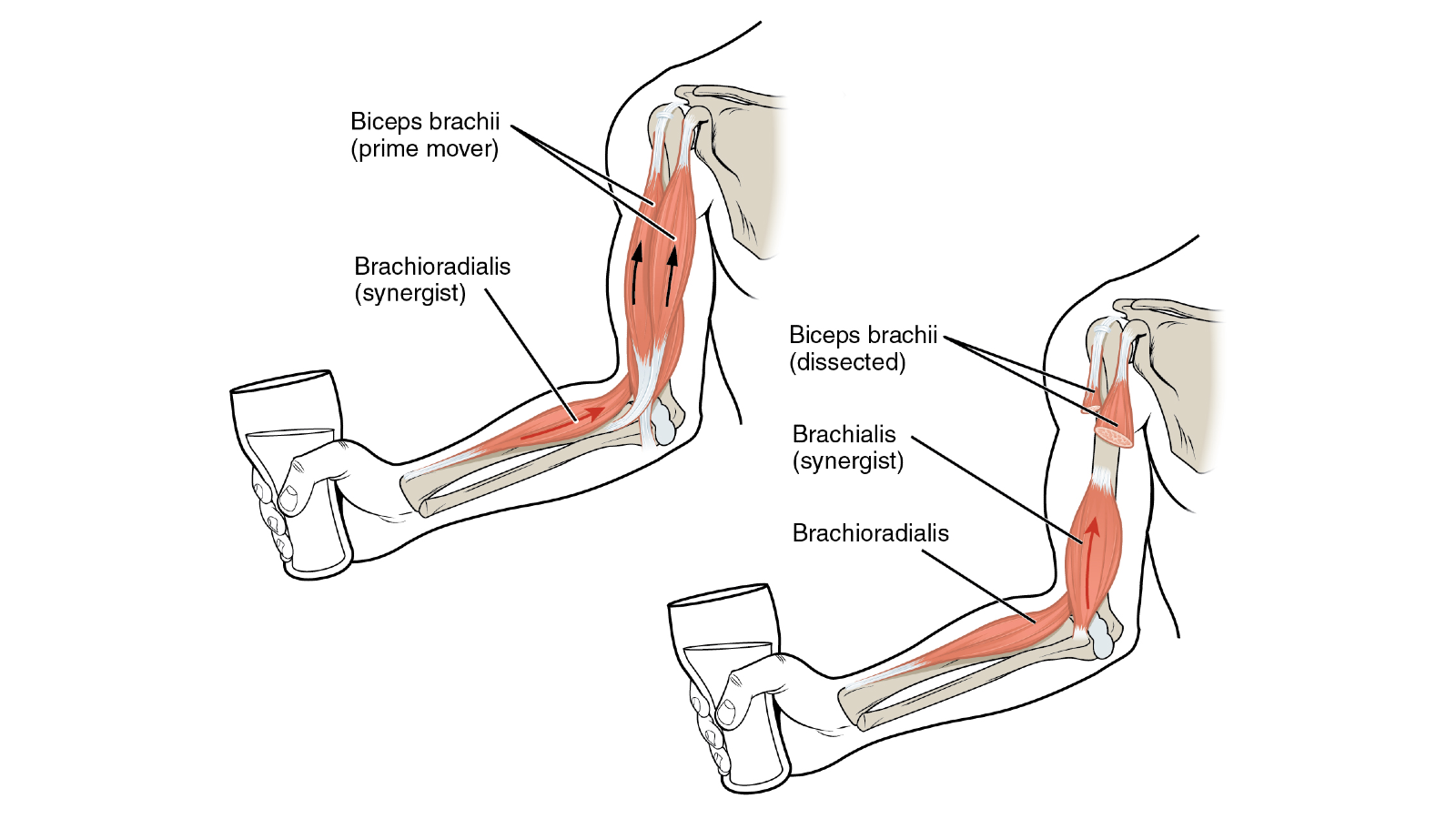
Synergist - Works together with other muscles such as agonists and antagonists to aid in the control of movement, an example of a synergist muscle is the muscles in the foot that help support the movement of the toes.
-
Fixator - Help to hold the body in place during movement, examples are the muscles in the ankle that keep the leg straight when walking.
The activation (contraction and relaxation) of muscles is controlled by a sensory reception process known as Proprioception. Specialised receptors (known as mechanoreceptors or proprioceptors) detect changes in the length of the muscle as a result of movement or changes in the surface, relaying information to the central nervous system. The signals sent to and from the spinal cord instruct the agonist muscle to contract. This is known as the stretch reflex.
Reciprocal inhibition uses the same sensory feedback system to cause an automatic relaxation of the opposing (antagonist) muscle to the one that is being stretched (agonist muscle). For example, when the bicep is flexed reciprocal inhibition sends a message, via the central nervous system, to relax the tricep which is the opposing muscle.
The different ways that muscles contract determine the way that they cause movements in the body. There are three main types of contractions:
-
Isotonic - Generate force through changes in the length of the muscle in the following two ways: Concentric (contractions that shorten the muscle) and Eccentric (contractions that lengthen the muscle). Squats, stair climbing, and bicep curls are examples of isotonic muscle contraction.
-
Isokinetic - Generate force through movements that are of a constant speed. Resistance based exercises that involve special equipment (like an exercise bike) allowing for variable resistance to a movement allowing for the muscle to contract at constant speeds is an example of isokinetic muscle contraction.
-
Isometric - Generate force without any change in the length of the muscle. Stationary exercises such as planks, wall sits, and bridges are examples of isometric muscle contraction.
The image below shows how a prime mover muscle biceps brachii is supported by a synergist muscle brachioradialis to perform the action of flexing the elbow joint to lift a glass.
The muscular system combined with the skeletal system (see below) forms the Musculoskeletal System. These two systems work in synchrony to enable locomotion of the body.
.jpg)
It is also an area that is prone to injury as a result of falls and trauma. In particular, injuries that affect the skeletal system include:
-
Sprains and Strains.
-
Ligament tearing.
-
Cramps, spasms, and inflammation.
Ensuring appropriate warm-up and cool-down activities are undertaken as well as wearing appropriate footwear, practising correct technique, and improving balance and coordination can help to prevent injuries to the musculoskeletal system.
There are two sections of the nervous system:
-
Central Nervous System (CNS) - Consisting of the brain and the spinal cord.
-
Peripheral Nervous System (PNS) - Consisting of individual nerve fibres (axons), thousands of nerve cells (neurons), and the sensory organs (eyes, nose, tongue, ears, and skin).
The central nervous system is the main component of the nervous system whilst the peripheral nervous system acts as a connecting 'highway' allowing the nerves to distribute messages between the body and the brain. These messages are known as nerve impulses.
The main functions of the nervous system are to:
-
Control the body's internal environment (homeostasis).
-
Facilitate memory and learning.
-
Control unconscious (autonomic) bodily functions such as heart rate, digestion, respiratory rate, pupillary response, urination, reflexes, and sexual arousal.
-
Initiate voluntary (somatic) actions, such as walking and talking.
The central nervous system is the main component of the nervous system whilst the peripheral nervous system acts as a connecting 'highway' allowing the nerves to distribute messages between the body and the brain. These messages are known as nerve impulses.
Nerve impulses are essentially a collection of electrically charged motor neurons (nerve cells) that conduct signals to pass information (messages) through the body and mobilise skeletal muscles. The combination of the motor neuron and the skeletal muscle fibres is known as a Motor Unit.
Motor Neurons are made up of:
-
Dendrites - The point of input for the nerve impulse.
-
Soma (or cell body) - Contains the nucleus which is the main part of the cell controlling its activity and containing the genetic material (chromosomes).
-
Axon (with an axon terminal) - The point of output for the nerve impulse.
The video below explains how the various components of the nervous system operate.
Skill-related components of fitness such as balance, agility, power and speed, reaction time, coordination, and proprioception all involve communication between the nervous system and the musculoskeletal system.
In regards to movement initiation, the nervous system is similar to a traffic highway, there are two lanes in which messages travel:
-
One direction consists of a series of afferent neurons (sensory nerve cells) connected by axons (nerve fibres) which distribute a message from a sensory point via a domino effect until it reaches the brain.
-
The other direction consists of a series of efferent neurons (motor nerve cells) connected by axons (nerve fibres) which distribute a message from the brain to the relevant muscle commanding it to move.

Involuntary movement (not controlled by the brain) is known as a reflex. A reflex is an autonomic (subconscious) response to stimuli. Reflexes assist in the function of maintaining homeostasis (a stable and relatively controlled internal environment) and include reactions such as breathing, blood pressure regulation (baroreceptor reflex), swallowing, sneezing, coughing and vomiting.
Reflex messages originate from the spinal cord or brainstem, thus the reaction time is much quicker than other movements. The circuit of message communication which controls reflexes is known as a reflex arc.
A stretch reflex (also known as a deep tendon reflex or myotatic reflex) is the response of the body when a muscle is passively stretched, causing the muscle to contract. It can be activated by external forces, for example when a doctor taps the patella tendon and causes your leg to jerk. The stretch reflex is also activated if the ground surface suddenly becomes uneven, helping to maintain balance. The purpose of a stretch reflex is to provide protection preventing muscle tearing due to sudden vigorous movement.
Proprioceptors (also known as mechanoreceptors) are the sensory receptors which help to activate the stretch reflex. They are primarily found in the muscles, tendons, and skin. The action of proprioceptors working to function muscles is known as proprioception.
The role of the nervous system in different types of training includes:
-
Recalling memorised information that relates to basic movement such as how to walk, run, or jump.
-
Learning how to undertake new exercise techniques by using sensory receptors such as eyes and ears to observe demonstrations and listen to instructions.
-
Sending messages between the brain and muscles to facilitate movement.
-
Adapting the body during exercise by accelerating heart rate, secreting adrenaline, and directing blood towards key muscles.
Whilst this is a complicated process it is also very fast. Nerve impulses take about seven milliseconds to complete the circuit between the sensory point the brain and the muscle. To put this speed into perspective there are 1000 milliseconds in 1 second.
Neuromuscular adaptations such as improvements in muscle reaction time can be made through regular practice as newly learned techniques are committed to memory and become easier and easier to undertake. This improves speed, agility, and coordination. Try routines such as exercise drills using agility ladders and cones that require rapid movements to maneuver between points or games involving catching balls to improve hand-eye coordination.
Another benefit of regular exercise on the nervous system is the boost of blood flow to the brain thus improving cognitive function. In particular, exercise has been found to help the following functions of the nervous system:
-
Mood Regulation - Exercise stimulates 'feel-good' neurotransmitters such as dopamine and serotonin and can improve mood disorders such as depression.
-
Anxiety - High-intensity aerobic activity has been shown to improve the possibility of panic attacks.
-
Focus and Attention - Additional blood flow and oxygenation improve the ability to focus and concentrate.
-
Sleep - Insomnia is aided by regular exercise, however, it is recommended to not engage in vigorous exercise late in the evening (within four hours of going to sleep) as this acts as an energiser.
-
Memory - Increased blood flow to the hippocampus (the brain's memory centre) has been found to improve conditions such as Alzheimer's Disease and Dementia.
This system essentially has two names, renal or urinary, and its main responsibility is to filter waste from the blood and produce urine as a means of expelling this waste. The main components of these systems are:
-
Kidneys - Two bean-shaped organs that are responsible for filtering and urine production.
-
Ureters - Two thin tubes that take urine from the kidney to the bladder.
-
Bladder - A sac that stores urine for a certain time until there is an opportunity to urinate.
-
Urethra - The tube that carries urine from the bladder as it exits the body.
In humans, as will most other animals, sexual reproduction is initiated to have babies. Essentially two cells, one from the male (sperm) and one from the female (ovum) must join together to create a baby (foetus). The various components of the reproductive system, which varies between men and women, are responsible for the production, distribution and/or protection/growth of these cells and any foetus that is produced as a result of reproduction.
There are three major parts of the respiratory system:
-
Airway - Includes the nose, mouth, pharynx, larynx, trachea, bronchi, and bronchioles and carries air between the lungs and the body’s exterior.
-
Lungs - Pyramid-shaped organs within the chest which bring oxygen into the body through the breathing process and expel carbon dioxide out.
-
Muscles of respiration - There are quite a few different muscles, however, the main ones are the diaphragm and the external intercostals. They help to expand the thoracic cavity to allow inhalation and compress it to allow exhalation.
The cells of the human body require a constant stream of oxygen to stay alive. The respiratory system provides oxygen to the body’s cells while removing carbon dioxide, a waste product that can be lethal if allowed to accumulate. This is known as exchange.
Respiratory or lung volume is the amount of air that can be held in the lungs and the amount of air that is inhaled and exhaled of the lungs. This volume amount has a resting level and also additional levels that can be utilised if need be.
During exercise, the muscles need more oxygen and they produce more carbon dioxide as a waste product. Therefore, the respiratory system (in conjunction with the circulatory/cardiovascular system) increases its volume level to supply extra oxygen.
There are four different levels of lung volume:
-
Tidal volume - The amount of air that can be inhaled or exhaled during a normal resting breath cycle, this is around 700ml - 1200ml of air for most people.
-
Inspiratory reserve volume - This is the amount of air that is able to be inhaled beyond the normal tidal volume. This is the action of taking a deep breath during exercise or when yawning. This is between 2000ml and 3000ml for different people.
-
Expiratory reserve volume - The amount of air that can be forcefully exhaled out after the tidal volume has been expelled (ie. exhaling with force).
-
Residual volume - Is the amount of air that is left in the lungs once the expiratory volume is forced out.
As fitness improves, the body becomes more efficient at getting oxygen into the bloodstream and transporting it to working muscles. Both aerobic and anaerobic exercise is beneficial to improving respiratory function. Aerobic activities such as walking, running and swimming improve the heart and lung capacity and anaerobic (strength training) improves core and respiratory muscle strength.
Some respiratory conditions such as asthma can be improved through regular exercise in particular routines that promote breathing techniques and building lung capacity such as yoga or swimming.
There are 206 bones in an adult human, which make up the framework and supporting structure of the body. Bones also protect the softer parts of the body such as the brain, nerves in the spinal cord, heart, lungs and liver. The muscular system interacts with the skeletal system to connect various bones and allow the body to move.
The skeleton is divided into two major sections:
-
Axial Skeleton - Runs along the body's vertical axis, supporting the head, neck, back and chest and providing protection for the vital organs. It is made up of 80 bones in the following regions. Skull, Hyoid, Auditory Ossicles, Ribs, Sternum, and Vertebral Column.
-
Appendicular Skeleton - Allows for movement and is what the skeletal muscles are attached to. Consists of 126 bones in the following regions: Upper limbs, Lower limbs, Pelvic girdle, Pectoral (shoulder) girdle.
.jpg)
The major bones of the body include:
-
Skull - Provides protection for the brain.
-
Shoulder Girdle - Connects the arms to the torso and allows for movements such as shrugging and lifting arms above the head.
-
Arms (humerus, ulna, and radius) - Allows reaching and pulling movements and connects to the hands.
-
Wrists and hands - The hands, in particular, allow for grasping and picking up of a objects.
-
Chest (Ribs) - Provides protection for the heart, lungs and liver.
-
Spine - Provides protection for the spinal cord, helps to maintain posture and allows twisting movements.
-
Pelvic Girdle - Provides protection for the reproductive, urinary, and digestive organs and also allows movement of the legs.
-
Legs (femur, tibia, and fibula) - Support the weight of the body and allows movements like walking and running.
-
Ankles and feet - Provide balance and movements like walking and running.
Each of these areas contains multiple smaller bones all contributing to either the axial or appendicular skeletons.
Where two bones meet is known as a joint. There are three different types of joints:
-
Synarthroses or Fibrous (immovable) - Two or more bones in close contact which have no movement. For example, the various bones (plates) of the skull.
-
Amphiarthroses or Cartilaginous (slightly moveable) - Two or more bones held tightly together and only capable of limited movement. For example, the vertebrae of the spine.
-
Diarthroses or Synovial (freely moveable) - The main joints responsible for functional movement, they are lubricated by synovial fluid to allow free movement.
Within the diarthrosis joint category, there are six types of freely moveable joints.
-
Ball and socket - Permits movement in all directions. For example, shoulder joint and the hip joint.
-
Hinge - Moves in one direction similar to a door opening and closing. For example, elbow joint and the knee joint.
-
Condyloid - Moves in a non-rotational manner. For example, finger joints and the jaw.
-
Pivot - Swivels in a ring formed from a second bone. For example, the ulna and radius bones that rotate the forearm.
-
Gliding - Provides limited movement by allowing bones to glide over one another in a smooth movement. For example, the wrist.
-
Saddle - Back and forth or side to side motion without full rotation. For example, the thumb.
In regards to the moveable joints, there is a further breakdown of how they operate known as the Lever System. A joint is an intersecting point between two or more bones, this joining point forms an axis (also known as a fulcrum) and the muscles apply force to move a weight (also known as the resistance). Lever systems are not limited to the human body, they exist in any situation where weight is being moved by a balancing force, examples in life include scissors, wheelbarrows, and shovels.
The lever system is divided into three different classes:
-
First Class - The fulcrum (axis) is positioned between the weight and the force (muscles). A real-life example of a first-class lever system is a pair of scissors, the fulcrum (axis) is the join of the blades, the weight (or resistance) is the paper or item being cut, and the force is the person's hand operating the handles. In the human body, first-class levers are not common, the main example of one is the atlantooccipital joint between the head and the first vertebrae. This joint is used for movements such as nodding of the head.
-
Second Class - The weight (or resistance) is positioned between the fulcrum (axis) and the force (muscles). A real-life example of a second-class lever system is a wheelbarrow, the weight is the item in the bed of the barrow, the fulcrum (axis) is the wheel, and the force is the hands of the person operating the handles. In the human body, an example of a second-class lever is the movement of the lower leg when an individual is standing on their toes.
-
Third Class - The fulcrum (axis) is positioned at the opposite end to the weight (or resistance) with the force (muscles) in the middle. A real-life example of a third-class lever system is someone using a fishing rod, the weight (a fish) is positioned at the end of the rod, the axis is one of the hands of the person holding the rod and the force is their other hand positioned mid-way down the rod. In the human body, an example of a third-class lever is the movement of the elbow joint when the force (muscles) pull on the bones of the forearm to lift the weight of the forearm, wrist, and hand with the axis being the elbow.
Bony Landmarks are grooves or prominences found on bones, that can be seen or felt externally (on a lean or muscular body). They can help to identify the location of other body structures, like muscles. The video below explains how bony landmarks can be identified. The context of the video is in relation to life drawing/painting the human body but the essential elements of anatomy are the same. Note: Because the context is in relation to art this video contains some scenes of discreet nudity.
The skeletal system combined with the muscular system forms the Musculoskeletal System. These two systems work in synchrony to enable locomotion of the body. It is also an area that is prone to injury as a result of falls, strains, and trauma.
In particular, injuries that affect the skeletal system include:
-
Broken bones.
-
Dislocation of joints.
Ensuring appropriate warm-up and cool-down activities are undertaken as well as wearing appropriate footwear, practising correct technique, and improving balance and coordination can help to prevent injuries to the musculoskeletal system.
A living thing that has an organised structure, can react to stimuli, reproduce, grow, adapt, and maintain homeostasis.Biology online
Essentially, the human body is an organism. We have an organised structure of cells, tissues, organs and organ systems and are capable of reacting to stimuli (via our senses), reproduce (via our reproductive systems), grow, adapt and maintain homestasis which means to have a stable and relatively constant internal environment (ie. our body temperature among other things). Organisms are not just limited to humans or other animals, they can exist on a much smaller scale, for example, fungi or bacteria.
Injury prevention is the practice of ensuring that any risks are known and identified so that fitness is able to be planned and instructed in a safe manner. Having a thorough understanding of human anatomy and physiology can help to you to anticipate, prevent, and if need be effectively treat injuries.
Consideration for injury prevention may include:
-
Conducting a thorough client history and fitness test to determine if there are any pre-existing health conditions or old injuries that may limit a client's ability to perform certain exercises or predispose them to risk of acute symptoms or injury.
-
Applying knowledge of human anatomy and physiology to adapt standard program plans to accommodate any risk factors associated with health conditions or injuries.
-
Incorporate appropriate intensity increases, rest / interval periods, and exercise variation to ensure over exertion is not experienced.
-
Ensuring appropriate warm-up and cool-down routines are conducted.
-
Provide appropriate monitoring and evaluation, especially for high-risk clients, being mindful of any prevalent symptoms or exercise intolerance signs.
In the unfortunate instance of an injury occurring it is essential that fitness professionals have a good understanding of first aid principles and techniques and also maintain connection with a range of medical and allied health professionals who can assist with the treatment of acute and also chronic conditions and injuries.