As a personal trainer, you must be confident in using terminology correctly so you can communicate with clients, colleagues, and other health professionals. Anatomical terms give you the language necessary to describe the functions, relationships, and purposes of anatomy and physiology.
In other words, knowing anatomical and physiological vocabulary helps you understand the body.
In this topic, you will learn:
- Anatomical position
- Directional terms
- Body planes and sections
- Movement terms
- Body cavities
- Homeostasis
- Human body systems
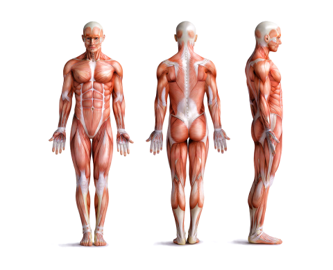
What is "Anatomy and Physiology"?
Studying two branches of science, anatomy and physiology, provides the foundation of knowledge of the body's parts and functions.
Anatomy is the study of the body's structures or parts. It asks...
What is that? And where is it in the body?
Physiology is the study of the functions of body parts. It asks...
What does it do?
Anatomy and physiology focus on different aspects of the study of the body, but they are typically studied together because the structure (anatomy) and function (physiology) of the body are closely related.
Structure mirrors function
As you progress with your anatomy and physiology studies, remember that structure mirrors function. Doing so will give you a deeper understanding of why and how something works, including:
- how and why muscles and joints move in particular ways.
- how organs function.
- how organs work together (organ systems).
- how the body works in sickness and in health.
Terminology checkpoint
The anatomical position (also known as the standard anatomical model), is the scientifically agreed-upon reference position for anatomical location terms. It is used to ensure a standardised approach to describing the position of appendages with respect to the body.
Why is it important?
Having a standard is necessary to avoid confusion since most organisms can take on many different positions that may change the relative placement of organs. This is of great importance in your role as a personal trainer for situations such as analysing postures, prescribing exercises, and designing programmes. Deviating from this can result in misdiagnosis and incorrect exercise prescription, causing potential risks to your clients and your reputation in the industry.
When talking about the locations of body parts, we must always consider the anatomical position.
How to describe the anatomical position to your clients
Use the following cues to keep your explanation simple.
- Stand upright
- Feet slightly apart
- Feet/toes pointing forward
- Palms facing forward
- Head facing forward with eyes looking ahead
Try it out
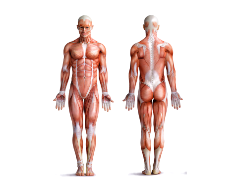
Practice standing in the anatomical position now, and describe it how you would to a client.
- Make sure your space is clear with room to move.
- Prop up your phone or recording device and take a video.
- Stand in the anatomical position.
- Describe the position using the suggested cues.
- Watch yourself back. Ask yourself if your demonstration is accurate and if your explanation is clear. Repeat the exercise until you can demonstrate and explain confidently.
Directional terms indicate the relation/location of one body part or organ to another. The anatomical position provides a standard for directional terms to be clearly defined worldwide. Under this standard, every organ or body part can be accurately and consistently described in relation to any other body part or organ.
How to use directional terms
When you use directional terms to describe an organ or structure, it is helpful if you give a reference point. For example, saying 'The kidney is inferior' doesn't tell us too much because we don't have a point of reference. We are left thinking, "inferior to what or where?" So, we need to build on this statement by saying, 'The kidneys are inferior to the diaphragm,' which gives us a clearer picture of their location, and that's the whole point of this additional terminology. We could build on this to pinpoint their location even more by saying, 'The kidneys are inferior to the diaphragm and anterior to the large intestines'.
Definitions
You will use directional terms regularly as a personal trainer. Start practising and becoming comfortable using them. You may find it helpful to keep a personalised terminology and vocabulary reference guide, either on paper or digitally.
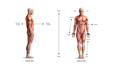
| Directional term | Definition | Example of use |
|---|---|---|
| Posterior | Nearer to or at the back of the body. | The esophagus (food tube) is posterior to the trachea (windpipe). |
| Anterior | Nearer to or at the front of the body. | The sternum (breastbone) is anterior to the heart. |
| Medial | Toward the midline of the body. Think of the spine as your 'plumb line' down the middle (midline) of the body, splitting your body into equal left and right halves. | The ulna is medial to the radius. |
| Lateral | Away from the midline of the body. | The lungs are lateral to the heart. |
| Superior | Toward the head (or towards the upper part of the body/structure). | The heart is superior to the urinary bladder. |
| Inferior | Toward the feet (or towards the lower part of the body/structure). | The stomach is inferior to the lungs. |
| Proximal | Toward the attached end of a limb or hollow tube/vessel. | The humerus (upper arm bone) is proximal to the radius (one of two forearm bones). |
| Distal | Away from the attached end of a limb or hollow tube/vessel. | The phalanges (finger bones) are distal to the carpals (wrist bones). |
| Superficial | Nearer to or on the surface. | The ribs are superficial to the lungs. |
| Deep | Farther away from the surface. | The ribs are deep to the skin of the chest and back. |
In your new language, you can now tell your friends: "the skin is superficial to the ribs, and the lungs are deep to the ribs".
Try it out
Can you use directional terms to accurately describe where these body parts are located?
Watch
Enjoy this light-hearted video showing the confusion your clients will have should they be bombarded with confusing scientific terminology. This video offers fun techniques to help remember these terms and what they mean, along with providing some handy ways to describe these to your clients.
Body planes (also known as planes of motion) divide the body or organs into different sections.
The planes of motion, plus the anatomical position, are the foundation of the terminology used by sports scientists and medical professionals to describe how the body (organs and/or cells) moves. Most exercises are performed predominately in one plane more than the others.
The planes
The body can be divided into 4 planes (3 main planes and an additional plane). There are other planes for deeper, more precise descriptions. However, in the fitness industry, using 3-4 planes is sufficient:
- Sagittal
- Frontal (coronal)
- Transverse
- Parasagittal
Every exercise can be related back to the movements we do in real life. We push, pull, flex, extend, squat, lunge, bend, and twist each and every day. Most exercises are predominately in one plane more than the others.
Imagine each plane as a plate of glass that cuts the body into either left and right (sagittal), front and back (frontal or coronal), or top and bottom (transverse) halves. Then imagine each of those plates of glass is a track that the body is moving on. If a movement seems to mostly track along one plate over the others, it can be classified as predominately in that plane of motion.
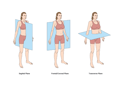
- The sagittal plane: The sagittal plane divides the body into equal left and right portions.
- The coronal or frontal plane: The coronal or frontal plane divides the body into posterior and anterior portions. Movement in this plane would be forward and backward movements of the whole body or limbs, such as arms and legs.
- The transverse plane: The transverse plane (also known as an axial plane or cross-section) divides the body into superior and inferior portions (think of the magician's assistant in the box!)
- The parasagittal plane: In addition to the sagittal plane, the parasagittal plane (not shown in the previous diagram) divides the body into unequal left and right portions (anything other than 50:50).
Transverse plane and rotation
Exercises in the transverse plane are rotational or anti-rotational movements. They can be:
- Spinal rotation
- Limb rotation
- Shoulder/hip rotation
The “plate of glass” analogy can be confusing when trying to understand the transverse plane. For spinal rotation, you may find it helpful to think of transverse plane movement in terms of an imaginary axis running vertically down through the centre of the head through the spine.
For limb rotation, any rotation inward or outward from your body is limb rotation and a transverse plane movement.
When your arm or leg is at 90 degrees to the body and moves away or towards the centre, this is shoulder or hip rotation and a transverse plane movement. Standing hip adduction happens in the frontal plane, but seated hip adduction occurs in the transverse plane.
Movement in 3D
Our bodies move in 3 dimensions (up/down, side-to-side, and front-to-back). Improving movement in all dimensions reduces the risk of injury and improves the chances of achieving fitness goals. To improve 3D movement, look at the body planes and choose exercises that move the body through all 3 planes of motion.

Try it out
Think about the planes as a track that your body moves along. When you squat, your body moves up and down along the sagittal plane. There is no purposeful movement side-to-side (along the frontal) or forwards and back (along the transverse). Therefore, the squat can be classified as a sagittal plane exercise (Payne, n.d.)
What is your favourite exercise that uses body weight only? I.e., no barbells, dumbbells, or machines. Perform it now if you can.
What body plane are you moving through, and what planes are you not moving through? How would you classify the movement?
Movement terms describe the common movements performed across joints due to muscle contractions. Learning these terms will help you break down the key movements in sporting and gym-based movements and help you identify the target muscles for each exercise.
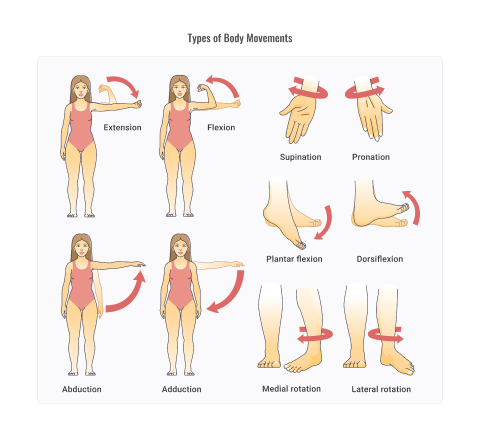
Movement terms can usually be paired up with counterparts, such as flexion and extension. This is because muscles tend to work in pairs to perform a movement. E.g., when the bicep muscle contracts, it shortens and pulls on the forearm bone, bringing it closer to the humerus (upper arm). To do this accurately and safely, the tricep muscle (posterior to the bicep) relaxes.
When a muscle contracts, it pulls on the bone, it does not push.
When you are discussing body movements, you need to provide a reference point. E.g., instead of saying, 'That is flexion', it would be more accurate and precise to name the body part/joint which is facilitating the movement. E.g., 'That is flexion (bending) of the knee'
Let's have a look at these movements:
- Flexion and extension
- Abduction and adduction
- Rotation
- Supination and pronation
- Supine and prone
- Lateral flexion
- Elevation and depression
- Protraction and retraction
- Ankle dorsiflexion and plantar flexion
Flexion and extension
These are common movements at most key joints. They are most often performed in the sagittal plane.
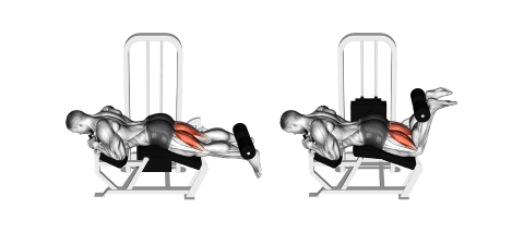
Flexion
Flexion describes a movement that causes a diminishing joint angle. It reduces the angle between the two bones in a joint, such as bending your elbow or knee. It can also refer to a joint movement that creates an angle between two bones at a joint that wasn't present in the anatomical position. E.g., bending your knee.
Extension
Extension refers to increasing a joint angle. It is essentially the opposite movement of flexion. E.g., straightening your knee or elbow after flexing it.
Hyper-extension
Hyper-extension refers to when the extension movement goes beyond normal parameters.
Try it now
Hold your arm in front of you, parallel to the ground, with your palm facing down.
Next, move your wrist into extension. Your palm is facing forwards, and your fingers pointing up. When your wrist is in extension, what happens to the angle between your palm and your forearm?
Now, move your wrist into flexion. Do your palm and fingers so your fingers are pointing down and your palm is facing back. Consider the angle between your forearm and palm now.
Abduction and adduction
These are common movements at many key joints. They are most often performed in the frontal plane.
Abduction
Abduction describes a movement away from the imaginary mid-line of the body (vertically). E.g., raising your arms at your sides, or the first leg movement during a star jump as you spread your legs apart. It usually refers to the limbs (arms and legs). It can also refer to the splaying of your fingers.
Adduction
Adduction describes a movement towards the imaginary mid-line of the body (vertically). E.g., lowering your arms to your sides after lifting them above your head, or the second leg movement during a star jump as you bring your legs back together. It can also refer to the bringing of your fingers back together after splaying them.
Horizontal abduction
Horizontal abduction occurs when you take your arms and legs away from the mid-line in a horizontal movement. E.g., the down phase of a chest fly or chest press.
Horizontal adduction
Horizontal addictions occur where, for example, you bring your arms and legs back towards the mid-line in a horizontal movement ('add-ing' the leg back towards the midline). E.g., the lifting/pushing phase of a chest fly or chest press.
Try it now
Stand in the anatomical position and move each limb through the abduction and adduction movements. Notice how their position to the mid-line changes with each movement.
Rotation
Rotation is a term used to describe movement around an axis. In the anatomical sense, this refers to rotation at a joint, or of the spine.
Rotation is typically described as external or internal. Key rotational aspects of the body:
- hip
- neck
- shoulder
- forearm (known as supination and pronation)
- spine
Try it now
Working from top to bottom, rotate the various joints, including your neck, shoulder, forearm, spine, and hip.
With each rotation, pay attention to where the axis is that the joint is rotating around.
Supination and pronation
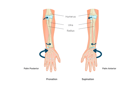
Supination is the movement that turns the palm of the hand upward towards the ceiling or sky, while pronation is the movement that turns the palm of the hand downward towards the floor or ground.
These movements are made possible by the two bones of the forearm, the radius and the ulna. The radius rotates around the ulna during supination, causing the palm to turn upward. During pronation, the radius rotates to its original position, causing the palm to turn downward.
Supination and pronation are important for many everyday activities, such as turning a doorknob, using a screwdriver, or throwing a ball. In the gym, trainers utilise these movements for grips during exercises. For instance, the client uses a supinated grip during a bicep curl, whereas during a bench press, they use a pronated grip.
Supine and prone
"Prone" refers to the body's position when it is lying face down, and "supine" refers to the body's position when it is lying face up.
In the prone position, the belly of the person is facing downwards, while the back is facing upwards. The head can be turned to one side or the other, and the arms can be positioned at the sides, or extended out in front of the body.
In the supine position, the belly of the person is facing upwards, while the back is facing downwards. The head and neck are usually in a neutral position, and the arms can be positioned at the sides, or extended out to the sides or overhead.
For example, a plank is a prone exercise, whereas sit-ups are done in a supine position.
Lateral flexion
This movement term describes a diminishing of the angle between the two structures of a joint but in the frontal plane. (Image 1 in the slider that follows.)
Elevation and depression
These terms refer to movements of the scapula. (Image 2 in the slider that follows.)
Protraction and retraction
These terms typically refer to the scapula (shoulder blade) and jaw. (Image 3 in the slider that follows.)
Ankle dorsiflexion and plantar flexion
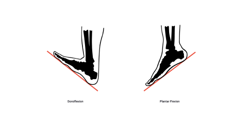
These terms refer to movements of the ankle. Remember that flexion is the diminishing angle between two bones in a joint.
Dorsiflexion
Dorsiflexion reduces the angle between the foot and tibia. This can be either when the toes are lifted towards the tibia (shin), or when the shin is rotated forward over the foot/toes (as in a lunge).
Plantar flexion
Plantar flexion increases the angle between the foot and tibia. Most often performed by pushing down the toes (as in a calf-raise). In other words, 'planting' your foot on the floor.
Try it now
Sit on a bed or the floor with your legs straight.
Flex your feet toward you, pushing the heel away and the toes forward to create dorsiflexion. Hold for 5 seconds.
Reverse the move, pointing your toes to create plantar flexion. Hold for 5 seconds.
Watch this interactive video tutorial on joint actions and have a go at the interactive activities.
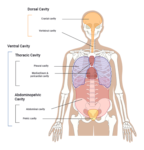
The body is composed of cavities. Cavities are hollow, fluid-filled spaces that encompass, protect, and provide a structure to hold organs in their place. They are separated from one another by bones, muscles, ligaments, and other structures.
Serous membrane
Each cavity is lined with its own serous membrane, a single-layered membrane that folds back on itself to produce a two-layered membrane structure. Within the membrane is a hollow space that is filled with serous fluid (a lubricating and shock-absorbing fluid produced by the cells of the membrane). The purpose of this fluid is to provide for smooth, frictionless movement of the two layers of the membrane when organs and body parts move and expand in size. It also provides an additional form of protection/shock absorption for the internal organs.
Function
The functions of body cavities are to:
- protect internal organs,
- support internal organs,
- provide space for organs to develop,
- and provide space for organs to move and expand (e.g., lungs, heart, and liver).
The ventral cavity
There are 3 main cavities within the torso, collectively making up the ventral cavity.
- The thoracic cavity
- The abdominal cavity
- The pelvic cavity
The thoracic cavity
Essentially the chest and everything it contains.
The main organs found in the thoracic cavity are:
- the lungs,
- the heart,
- and the diaphragm.
The abdominal cavity
This is the area under your abdominals (from your diaphragm down to the top of your pelvis).
It contains:
- the upper digestive organs,
- the spleen,
- the kidneys,
- and the ureters (tubes that deliver urine from the kidneys to the bladder).
Pelvic cavity
This cavity covers the area from the top of your pelvis to your sexual organs.
It includes:
- the urinary bladder,
- the urethra,
- and the reproductive organs (testes, ovaries).
Try it out
Can you identify where the following cavities are on your body?
- Dorsal cavity, including the cranial cavity and the vertebral cavity
- Thoracic cavity, including the pleural cavity and the mediastinum and pericardial cavity
- Abdominal cavity
- Pelvic cavity
Watch
Watch the video, which puts an everyday spin on cavities and the terminology that is associated with their physiology and function. This video offers some fun ways to view our 'squishy meat sack!' You don't need to learn everything in this video but do become familiar with the terms and the information.
Homeostasis is the reason why the body does what it does, when it does it, and how it adapts to finding new ways to continue doing what it does.
It is responsible for maintaining balance and equilibrium in the body as a whole.
Homeostasis is the condition of equilibrium of the body’s internal environment due to the constant interaction of the body’s many regulatory processes, and changing external environment.
Homeostasis is a dynamic condition which means it never sleeps. It constantly works to maintain our internal environment in its 'optimal' or most functional state.
Homeostasis can be defined as:
maintaining the body’s internal environment, in response to changing conditions, within normal bodily limits.
Body temperature
Let's look at body temperature as an example of how homeostasis works.
Normal body temperature ranges from 36.5 to 37.5 degrees Celsius, with a fever setting in at approximately 37.6 degrees. This shows us the very small window of one degree that the body and the relevant systems coordinate to maintain.
If you have ever had a fever, you will know how uncomfortable you felt! In some cases, patients are admitted to the hospital. Mild and moderate fevers cause weakness and exhaustion. A very high fever (40 degrees or higher) can result in convulsions and death. This is just one example of factors within the body that is constantly under review and stabilised. This is like a continuous, 24/7 risk analysis project management of our bodies!
Environmental impacts
Homeostasis is constantly being challenged and disturbed every second of every day. Consider the following two situations.
- You take a walk outside where it is warm, and you become out of breath and inhale the air from the surrounding environment.
- In another situation, you are sitting at your desk, slouched over, tired, with dry eyes.
How does your body react to different environments?
Every moment of the day and night your body and its systems are constantly:
- surveying the body's internal systems.
- communicating with other cells and systems of the body to let them know what is happening.
- recruiting additional cellular or system-based support when it is needed.
There are 11 systems in the human body. The systems work independently of each other, but if one is not functioning as intended, the others will try to correct the problem. The systems work together to maintain homeostasis.
Main systems of the human body:
- Cardiovascular
- Digestive
- Endocrine
- Integumentary
- Lymphatic
- Muscular
- Nervous
- Reproductive
- Respiratory
- Skeletal
- Urinary
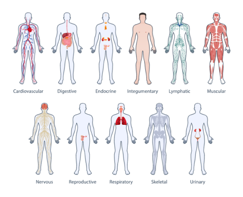
Human Body Systems by OpenMD.com, © OpenMD.com
Strengthen your learning by completing the practical activity.
Before moving to the next topic, check that you have a good grasp of anatomical terminology.
Watch
For a fun overview, which contains a little extra detail, enjoy the following video.
