In this section you will learn to:
- Obtain information regarding the physical health status of the person, through observation, questioning and interpretation of documentation.
- Use and interpret health terminology that describes the normal structure, function, and location of the major body systems.
- Use and interpret information that relates to the interrelationships between major components of each body system and other structures.
- Recognise variations from normal health status in consultation with Allied Health Professional.
- Report variations to Allied Health Professional in accordance with organisational policies and procedures.
Supplementary materials relevant to this section:
- Reading B: Patient-clinician communication in hospitals
- Reading C: Heimlich Maneuver
- Reading D: An overview and management of osteoporosis
- Reading E: Full Body Stretching Routine
- Reading F: The Stroke Survivor
- Reading G: Medical Assistance in the War Against Pathogens
- Reading H: Convulsions in Children with Covid-19
In the previous section of this module, you were given a set of guidelines to follow when it came to having tasks delegated at you. In this section of the module we are going to look at obtaining information regarding the patient’s health status such as their basic information like this height and weight for example and moving towards more detailed information such as their therapeutic remedies that have been prescribed by an Allied Health Professional.

Documentation is essential to maintain safe, high-quality care in the health sector. It is used:
- as a communication tool to facilitate the continuum of patient/client care
- to allow evaluation of the care provided
- for research or epidemiological needs
- to meet statutory requirements

Remember that as an AHA you need to document significant aspects of patient/client care to keep yourself safe from any breach in policy including:
- all direct contact with the patient/client, carers or other related individuals and any other significant activity that relates to patient/client care (including indirect contact), for example, missed or cancelled appointments, information provided/posted to the patient/client.
Principles of documentation

The format of your entries will be guided by Hospital and Health Service (HHS) policy as well as discipline and work unit-specific practices depending on the organisation you are employed at. Regardless of the format, the following principles of documentation will be applicable:
- always document as soon as possible after the intervention (e.g., occasion of service, phone call)
- content should be concise, relevant, appropriate and accurate
- do not diagnose
- use only standard abbreviations and avoid non-standard terminology
- it is important that your documentation can be understood by anyone reading the health record
- check with your supervisor regarding which abbreviations you can use
- if you don’t recognise abbreviations you see in other entries, ask your supervisor or another allied health professional to explain these to you. The table below depicts some commonly used abbreviations that you may come across when it comes to working in the Allied Health industry.
| Documentation format | Time descriptors | ||
|---|---|---|---|
| S/ | Subjective information | mane | In the morning |
| O/ | Objective information | am | morning |
| I/ | Intervention | pm | Afternoon |
| Rx | Treatment | 1/7 | One day |
| A/ | Assessment | 1/52 | One week |
| P/ | Plan | 1/12 | One month |
| c/o | Complains of | Therapy-specific | |
| o/e | Objective Examination | Mob | Mobilise |
| // | Outcome of intervention | ROM | Range of motion |
| Mobility aids | HEP | Home exercise program | |
| SPS | Single point stick | ex | Exercise |
| 4PS | Four-point stick | ADL | Activities of daily living |
| 4WW | Four wheeled walker or 'wheelie walker' | Activities of daily living (ADL) aids | |
| FASF / ESF | Forearm support frame or Elbow support frame | OTF | Over toilet frame |
| W/C | Wheelchair | HHH | Hand held hose |
| Indep | Independent | Other | |
| Assistance required | Pt | Patient | |
| s/v | Supervision | WNL | Within normal limits |
| Min | Minimal | NAD | Nil abnormality detected |
| Mod | Moderate | Physical status | |
| Max | Maximal | BMI | Body mass index |
| 1xA | One person assist | Wt | Weight |
| 2xA | Two person assist | ||
- be objective and factual
- be specific and avoid general terms.
- objective information is what is directly seen, heard, felt, or smelled:
- seen—for example, recording observations regarding bleeding, deformities, drainage, colour of urine, patient/ client posture and/or attitude.
- heard—for example, the patient/client’s comments, moaning, breathing abnormalities, speech sound errors.
- smelled—for example, vomitus odour.
- felt—for example, hot, cold, dry or moist skin, range of movement.
- subjective information is your own personal bias, judgement or speculation about the patient
- subjective statements should be avoided, that is, do not record your own emotional statements or moral judgements.
- if you think it is important to include a subjective statement made by the patient/client or another person you can record this (e.g., ‘husband reports improved speech’).
Always remember it is your responsibility to understand and clarify any information regarding the policies and procedures behind documenting patient information at the organisation you are employed at.
Tip - Find out more
The Administrative Record Keeping Guidelines from the Australian Department of Health have been designed to provide you with a range of helpful suggestions, general information and guidance to assist you to implement and maintain adequate record keeping systems that you may find beneficial for your practice.

As an AHA you will be required to gather information about a patient’s health condition, which can be done through observation, questioning and interpretation of documentation such as a referral from their doctor outside the service. Let’s take a look at how you are required to consensually obtain information from a patient.
You must only collect health information by lawful and fair means.
- ‘Lawful’ collection is a collection that does not breach any State, Territory or Commonwealth law.
- ‘Fair means’ is collecting without intimidation or deception, and in a way that is not unreasonably intrusive.
Under the Telecommunications (Interception) Act 1979 (Cth) and State and Territory listening devices laws, it is illegal to record a telephone consultation without informing the patient the call is being recorded. Collection via this method would therefore not be by lawful means. If a call is to be recorded or monitored, you must inform the individual at the beginning of the conversation so that the individual has a chance to end the call or ask not to be recorded.
Patients may be concerned or embarrassed about discussing health issues in an open or public area such as a waiting room or open pharmacy. When collecting health information, you should consider the surroundings and take additional steps where required to make the patient more comfortable. For example, you might lower your voice so only the patient can hear what you are saying, take the patient to one side, or use a private room.
(Australian Government, 2019)
The following steps will support you in making sure you gather information from a patient appropriately,

- Observation:
- You can observe or visually see the person's physical appearance, demeanour, and posture. Look for any visible signs of distress, discomfort, or abnormalities such as difficulty breathing, unusual coloration, swelling, or mobility issues.
- Try and pay attention to the person's gait, balance, and coordination as they move or walk.
- You can observe any physical limitations or restrictions in movement, such as the use of mobility aids or braces.
- Try and notice any non-verbal cues, such as facial expressions, gestures, or signs of pain or discomfort.
- Questioning:
- You can engage in open-ended and specific questioning to gather information about the person's physical health. Ask about any current or past medical conditions, recent injuries or surgeries, chronic pain or discomfort, and any symptoms they may be experiencing.
- Try and inquire about the person's ability to perform activities of daily living, such as bathing, dressing, eating, and mobility.
- You can ask about their current medications, including dosage and frequency, as well as any allergies or adverse reactions to medications.
- Interpretation of documentation:
- Try and review any existing medical records, reports, or documentation related to the person's physical health. This may include physician notes, diagnostic test results, rehabilitation plans, and progress reports.
- You must interpret the information within the documentation, paying attention to any pertinent details regarding the person's physical health status, diagnoses, treatments, and prescribed interventions.
- Remember as an AHA to always take note of any precautions, limitations, or contraindications mentioned in the documentation that may impact the person's physical health or the activities they can engage in.
- Collaboration with healthcare professionals:
- Make sure you consult with the healthcare professionals involved in the person's care, such as physicians, nurses, or therapists, to gather additional information or seek clarification on specific aspects of the person's physical health.
- As an AHA you must collaborate with the healthcare team to ensure a comprehensive understanding of the person's physical health status, including any recent changes, updates, or recommendations for their care.
- Documentation and reporting:
- You must maintain accurate and detailed records of your observations, the information gathered through questioning, and any interpretation of documentation.
- You should report any significant findings, concerns, or changes in the person's physical health status to the appropriate healthcare professionals or your supervisor, following the established communication protocols within your organisation.
Remember to always adhere to privacy and confidentiality regulations when accessing and sharing information about an individual's physical health. Obtain proper consent and ensure that sensitive information is handled securely and shared only with authorised individuals involved in the person's care.
Read
Reading B: Patient-clinician communication in hospitals
Reading B describes how workers in the health care sector can communicate effectively through two-way communication (spoken, written and non-verbal) that engages patients in decision making and care planning. It is tailored, open, honest and respectful and there is an opportunity for clarification and feedback.

The basic building block of living organisms is the cell. The human body is composed of countless millions of cells. There are many different types of cells, which are specialised so they can carry out functions in the body. Despite these differences, certain features are the same in most cells. The figure below shows the structures present in a typical animal cell.

The living material that makes up a cell is called cytoplasm. It has a texture rather like sloppy jelly, somewhere between a solid and a liquid.
The largest organelle in the cell is the nucleus. Nearly all cells have a nucleus, with a few exceptions such as red blood cells. The nucleus controls the activities of the cell. It contains chromosomes (46 in human cells) which carry the genetic material or genes. Genes control the activities in the cell by determining which proteins the cell can make (see below). One very important group of proteins found in cells is enzymes. Enzymes control chemical reactions that take place in the cytoplasm.
All cells are surrounded by a cell membrane, sometimes called the cell surface membrane to distinguish it from other membranes inside the cell. This is a thin layer like a ‘skin’ on the surface of the cell. It forms a boundary between the cytoplasm of the cell and the outside.
However, it is not a complete barrier. In fact, as you will see, the membrane can actively control the movement of some substances.
One organelle that is found in the cytoplasm of nearly all living cells is the mitochondrion (plural mitochondria). In cells that need a lot of energy, such as muscle or nerve cells, there are many mitochondria. This gives us a clue as to their role. They perform some of the reactions of respiration, releasing energy that the cell can use. Most of the energy from respiration is released in the mitochondria.
Differentiation of cells

A human begins life as a zygote, which divides to form two cells, then four, then eight and so on, until an embryo is formed, containing many millions of cells.

As the developing embryo grows, cells become specialised to perform particular roles. This specialisation is also under the control of the genes and is called differentiation. Different kinds of cell develop depending on where they are located in the embryo, for example, a nerve cell in the spinal cord, or an epidermal cell in the outer layer of the skin.
Muscle Tissues

Muscle tissue consists of cells that are specialised to shorten, or contract, resulting in movement of some kind. Muscle tissue is composed of tightly packed cells called muscle fibres. The fibres are generally long and thin and aligned parallel to each other. Muscle fibre contains proteins which interact to make the cell contract. There are three types of muscle tissue: voluntary, involuntary and, cardiac. They vary somewhat in body location, structure, and function, but they all do essentially the same thing— when stimulated, they contract. For now, we’ll focus solely on differences between the three types of muscle tissue.
Skeletal muscle tissue (voluntary)
Skeletal muscle tissue connects to tendons, which attach to bones. When skeletal muscles contract, they cause body parts to move. A skeletal muscle may contain thousands of individual cells, all aligned parallel to each other. A skeletal muscle is called voluntary muscle because we can exert conscious control over its activity. Skeletal muscle cells are activated only by nerves.
Smooth muscle tissue (involuntary)
These tissues are hollow organs and tubes, including blood vessels, digestive tract, uterus, and bladder. These slim cells are much smaller than skeletal muscle cells and have only one nucleus, like cardiac muscle. The cells are aligned roughly parallel to each other. In blood vessels, they are generally aligned in a circular fashion around the vessel. When smooth muscle cells shorten, the diameter of the blood vessel is reduced. Smooth muscle cells taper at both ends, and there are gap junctions between adjacent cells so that when one contracts, nearby cells also contract. Like cardiac muscle, smooth muscle is involuntary in that we cannot control its contractions consciously.
Cardiac muscle tissue
Cardiac muscle tissue (in Greek, kardia means “the heart”) is found only in the heart. The individual cells are much shorter than skeletal muscle fibres, and they have only one nucleus. Like skeletal muscle, the cells are arranged parallel to each other. Cardiac muscle cells are short and blunt ended, with gap junctions between the ends of adjoining cells. The gap junctions represent direct electrical connections between adjoining cells, so when one cell is activated, it activates it neighbours down the line. Because of these gap junctions, the entire heart contracts in a coordinated fashion. Cardiac muscle is considered involuntary because the heart can contract rhythmically entirely on its own, without any conscious thought on our part and without any stimulation by nerves.
Tip - Remember:
Skeletal muscle tissue connects to tendons and bones. Smooth muscle tissue surrounds organs and tubes. Cardiac muscle tissue comprises most of the heart. The common feature of all muscle tissues is that they contract, producing movement
Other tissues include blood, which is made of various types of red and white blood cells in a liquid matrix called plasma, and nervous tissue, which makes up the brain, spinal cord, and nerves. A collection of different tissues carrying out a particular function is called an organ. The main organs of the human body are shown in the image below.


The human body is a very complex machine. It needs food for growth and repair of tissues, as well as for a supply of energy. In this section, we look at how the cells and tissues we spoke about above are organised into organs and organ systems that comprise your body, as well as how they work together to maintain the health and stability of your body.
Many of the more complex functions of multicellular organisms (such as pumping blood or digesting food) cannot be carried out by one tissue type alone. Organs are structures composed of two or more tissue types joined together that perform a specific function or functions. Your heart is an organ because the tissues that comprise it share one essential function: They pump blood. Most of the heart consists of cardiac muscle, but there is also smooth muscle in the blood vessels that supply the cardiac muscle. The heart contains nervous tissue that affects the rate at which the organ beats. Some connective tissue, primarily in the heart valves, open and close to control blood flow within the heart, and even a thin layer of epithelial tissue lines the heart chambers. Some organs have several functions. For example, the kidneys remove waste and helps control blood pressure.
Organ systems are groups of organs that together serve a broad function that is important to survival either of the individual organism or a species. For example, survival of an individual organism depends on respiration, movement, or excretion of wastes, and survival of a species relies on reproduction. A good example is the organ system responsible for the digestion of food. Your digestive system includes your mouth, throat, esophagus, stomach, intestines, and even your liver, pancreas, and gallbladder. All these organs must interact and be controlled and coordinated to accomplish their overall function.
(Johnson, 2017)

Cells get their energy by oxidising foods such as glucose. This process is called respiration and needs a continuous supply of oxygen from the blood. In addition, the waste product carbon dioxide needs to be removed from the body. These gases are exchanged between the blood and the air in the lungs. In this section, you will learn about the processes of respiration and gas exchange.
Tip - Remember:
Respiration is called an oxidation reaction because oxygen is used to break down food molecules
The organs of the respiratory system, which are located in the chest cavity, ensure that oxygen enters the body and carbon dioxide leaves the body. During inspiration, or inhalation (breathing in), air is conducted from the atmosphere to the lungs by a series of cavities, tubes, and openings. During expiration, or exhalation (breathing out), air is conducted from the lungs to the atmosphere by way of the same structures.
The respiratory tract extends from the nose to the lungs. Note the organs in the upper respiratory tract and the ones in the lower respiratory tract.

The upper respiratory tract

The nasal cavities, pharynx, and larynx are the organs of the upper respiratory tract.
The nose
The nose opens at the nares (nostrils) that lead to the nasal cavities. The nasal cavities are narrow canals separated from each other by a septum composed of bone and cartilage. Air entering the nasal cavities encounters large, stiff hairs that act as a screening mechanism. The hairs filter the air and trap small particles (dust, mould spores, pollen, etc.) so that they don’t enter air passages. The rest of the nasal cavities are lined by mucous membrane.

Tip - Did you know that?
Scientists have found that air travels at 100 miles an hour during a sneeze. This is enough force to propel sneeze droplets up to 12 ft away from the person sneezing. If a sneeze is stifled, the air is forced into the auditory (eustachian) tube and middle ear, potentially causing damage to the middle ear.
The pharynx
The pharynx, commonly referred to as the throat, is a funnel shaped passageway that connects the nasal and oral cavities to the larynx. The pharynx has three parts: the nasopharynx, where the nasal cavities open above the soft palate; the oropharynx, where the oral cavity opens; and the laryngopharynx, which opens into the larynx.
The larynx
The larynx is normally open, allowing air to pass, but the oesophagus is normally closed and opens only when a person swallows. If someone swallows and some of the food enters the larynx, coughing occurs in an effort to dislodge the food. If the passageway remains blocked, the Heimlich manoeuvre can be used to dislodge food blocking the airway.
Read
Reading C: Heimlich Manoeuvre
Reading C explains the steps taken in a Heimlich Manoeuvre, which is a rapid first-aid procedure to treat choking due to upper airway obstruction by a foreign object
When a person’s air supply is partly or fully blocked, they are at risk of injury and death from choking or suffocation. This can occur when breathing is disrupted by a physical blockage, such as an object in the windpipe, or when a person is trapped in a low oxygen environment.
Choking and suffocation injuries resulted in 1700 hospitalisations between 2020 and 2021, which is 6.5% per 100,000 population.
This represents 7% of injury deaths and 0.3% of injury hospitalisations.
Choking on objects is the most common cause of death in this category, while a foreign body in the respiratory tract is the most common cause of hospitalised injury.
Older people have the highest rates of death by choking and suffocation, while young children have the highest rates of hospitalisation.
Rates of death due to choking and suffocation were highest for people aged sixty-five and over, while young children aged 0 to 4 had the highest rates of hospitalisation compared with other life-stage age groups.
Of the hospitalisations in 2020–21:
Thirty-two percent were for people aged 65 and over
26% were for children aged 0–4.
Males were 1.3 times as likely as females to be hospitalised for choking or suffocation in 2020–21.
Of the deaths in 2019–20, 80% were for people aged 65 and over.

The lower respiratory tract

Once the incoming air makes its way past the larynx, it enters the lower respiratory tract. The lower respiratory tract consists of the trachea, the bronchial tree, and the lungs.
The trachea
The trachea, commonly called the windpipe, is a tube connecting the larynx to the primary bronchi. It is separated from the oesophagus by a flexible, muscular wall. This orientation allows the oesophagus to expand when swallowing. The goblet cells produce mucus, which traps debris in the air as it passes through the trachea. The mucus is then swept toward the pharynx and away from the lungs by the cilia that project from the epithelium. When you cough, the tracheal wall contracts, narrowing its diameter.
Therefore, coughing causes air to move more rapidly through the trachea, helping expel mucus and foreign objects. Smoking is known to destroy the cilia; consequently, the soot in cigarette smoke collects in the lungs. Smokers often develop heavy coughs as a result. If the trachea is blocked because of illness or the accidental swallowing of a foreign object, a breathing tube can be inserted by way of an incision made in the trachea. This tube acts as an artificial air intake and exhaust duct. The operation is called a tracheostomy.
Tobacco smoking is one of the largest preventable causes of death and disease in Australia. Smoking is estimated to kill almost 20,500 Australians a year (13% of all deaths) and was responsible for 8.6% of the total burden of disease in Australia in 2018. It is associated with an increased risk of health conditions including heart disease, diabetes, stroke, cancer, kidney disease, eye disease and respiratory conditions such as asthma, emphysema, and bronchitis.
In 2020-21, one in ten people (10.7% or 2.1 million people) aged 18 years and over were current daily smokers, with men more likely than women to smoke daily (12.6% compared to 8.8%). One in twelve (8.3%) people aged 18-24 years smoked daily, and this increased with age until 55-64 years where the rate peaked at 13.7%.

The bronchial tree (bronchi/bronchioles)
The trachea divides into right and left primary bronchi (singular term - bronchus), which lead into the right and left lungs. The bronchi branch into a few secondary bronchi that also branch, until the branches become bronchioles, which are about 1 mm in diameter. The bronchi resemble the trachea in structure. As the bronchial tubes divide and subdivide, their walls become thinner, and the small rings of cartilage are no longer present.
During an asthma attack, the smooth muscle of the bronchiole’s contracts, causing bronchiolar constriction and characteristic wheezing. Each bronchiole leads to an elongated space enclosed by a multitude of air pockets, or sacs, called alveoli.
Asthma is a long-term lung condition that is caused by narrowing of the airways when they become inflamed [1]. People with asthma have trouble breathing, and the most common symptoms are wheezing, coughing, breathlessness, and chest tightness. Asthma may affect people of all ages and can usually be managed through treatment such as medication use (e.g., reliever inhalers and preventer/controller medications) and a healthy lifestyle.
Just under 2.7 million people (10.7% of the total population) had asthma in 2020-21. Females were more likely than males to have asthma (12.0% compared to 9.4%); however, the rate of asthma was similar in children aged 0-14 years (9.5% and 7.9%).

The lungs
The lungs are paired, cone-shaped organs in the thoracic cavity. In the centre of the thoracic cavity are the trachea, heart, thymus, and oesophagus. The lungs are on each side of the trachea. The right lung has three lobes, and the left lung has two lobes, allowing room for the heart, which points left. Each lobe is further divided into lobules, and each lobule has a bronchiole serving many alveoli.
Pneumonia is a common and potentially fatal lung infection that should not be underestimated. It can be caused by viruses, bacteria, or fungi. When you breathe, air travels through the lungs to the alveoli or air sacs. If you have pneumonia the air sacs in your lungs fill with secretions and fluids that obstruct normal air flow. There are many types of pneumonia, one of the most common and life-threatening types is pneumococcal pneumonia, which is caused by the bacterium, Streptococcus pneumoniae.
This bacterium is responsible for approximately 1.6 million deaths each year world-wide. Healthy people may carry it in their nose and throat. While most of the time this does not cause any illness, vulnerable groups such as those over the age of 70, infants and people with a chronic illness may develop pneumococcal disease.
(Lung Foundation Australia, n.d.)
Tip - Remember:
The thin layer of fluid lining the inside of the alveoli comes from the blood. The capillaries and cells of the alveolar wall are ‘leaky’, and the blood pressure pushes fluid out from the blood plasma into the alveolus. Oxygen dissolves in this moist surface before it passes through the alveolar wall into the blood. If you want to know more about pneumonia clicks on the link - https://www.youtube.com/watch?v=JxZBbuaPxJk&t=25s
Mechanism of Breathing (The basics of breathing in and out)

Ventilation means moving air in and out of the lungs. This requires a difference in air pressure – the air moves from a place where the pressure is high to one where it is low. Ventilation depends on the fact that the thorax is an airtight cavity. When we breathe, we change the volume of our thorax, which alters the pressure inside it. This causes air to move in or out of the lungs. There are two movements that bring about ventilation, those of the ribs and the diaphragm.
If you put your hands on your chest and breathe in deeply, you can feel your ribs move upwards and outwards. They are moved by the intercostal muscles. The outer (external) intercostals contract, pulling the ribs up. At the same time, the muscles of the diaphragm contract, pulling the diaphragm down into a more flattened shape. Both these movements increase the volume of the chest cavity and cause a slight drop in pressure inside the thorax compared with the air pressure outside. Air then enters the lungs (inhalation or inspiration).
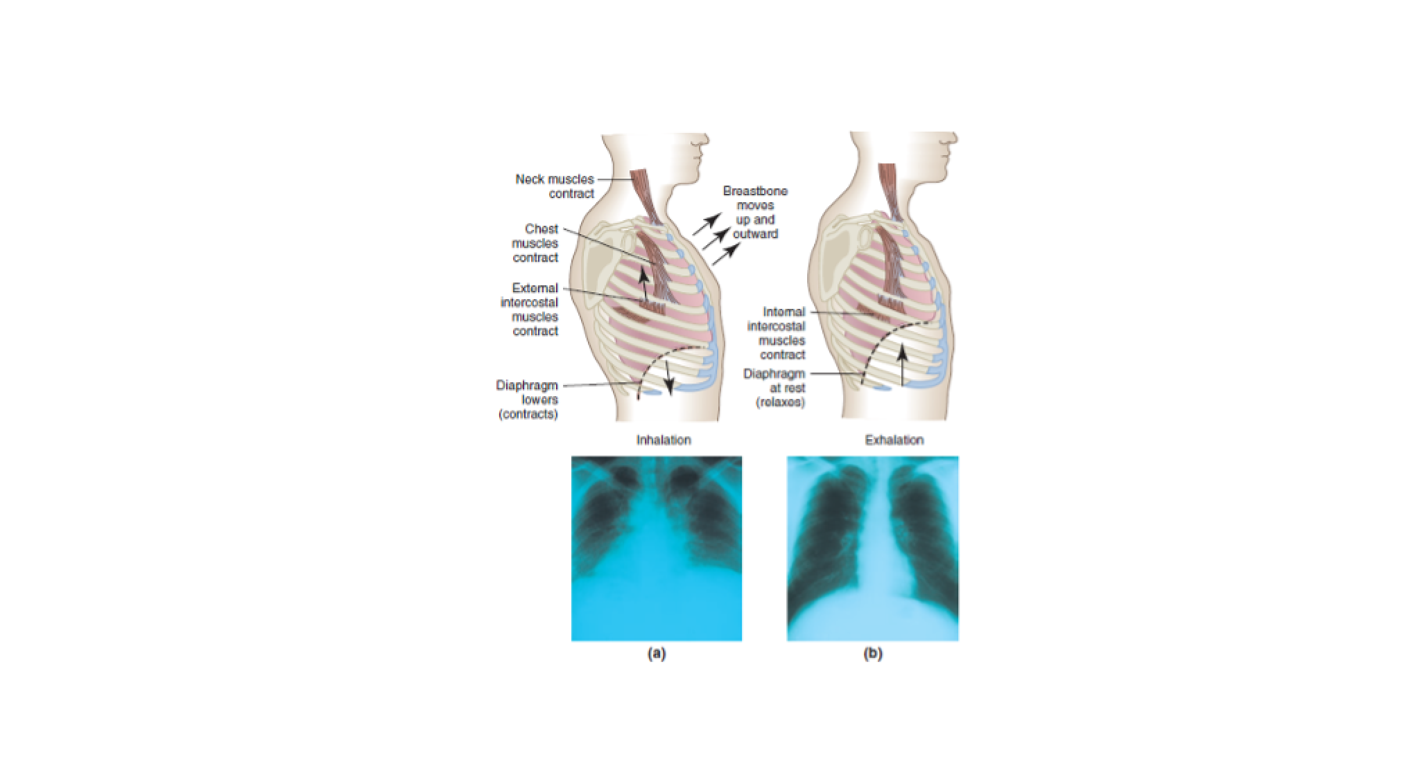
The opposite happens when you breathe out deeply. The external intercostals relax and the internal intercostals contract, pulling the ribs down and in. At the same time, the diaphragm muscles relax, and the diaphragm goes back to its normal dome shape. The volume of the thorax decreases and the pressure in the thorax is raised slightly above atmospheric pressure. This time, the difference in pressure forces air out of the lungs. Exhalation (expiration) is helped by the fact that the lungs are elastic, so they tend to collapse and empty like a balloon.
Changes in the position of the ribs and diaphragm during breathing.
- breathing in (inhalation)
- breathing out (exhalation)
It is important that you remember the changes in volume and pressure during ventilation. If you have trouble understanding them, think of what happens when you use a bicycle pump. If you push the pump handle, the air in the pump is squashed, its pressure rises, and it is forced out of the pump. If you pull on the handle, the air pressure inside the pump falls a little and air is drawn in from outside. This is similar to what happens in the lungs.
Read
Reading D: Imbalanced Breathing in Connection with Health Problems
Reading D describe the most common direct or indirect connections between imbalanced breathing and health. Imbalanced breathing potentiates symptoms, thus perpetuating the problematic situation. This is very common in states of overarousal, such as stress, anxiety, and pain. In addition, through stress reactions, conditions such as physical illness or surgery may contribute to the development of imbalanced breathing patterns and aggravation of symptoms.
Gas exchange
You can tell what is happening during gas exchange if you compare the amounts of different gases in atmospheric (inhaled) air with the air breathed out. The table below shows the approximate percentage volume of gases in inhaled (atmospheric) and exhaled air. Exhaled air is also warmer than inhaled (atmospheric) air and is saturated with water vapour.
| Gas | Inhaled (atmospheric) air | Exhaled air |
|---|---|---|
| Nitrogen | 78 | 79 |
| Oxygen | 21 | 16 |
| Carbon dioxide | 0.04 | 4 |
| Other gases | 1 | 1 |
Read
Reading E: Upper Respiratory Tract Infection
Reading E examines when an upper respiratory tract infection should be considered on differential diagnosis and how to properly evaluate it.

Humans are vertebrates – the human body is supported by an internal skeleton made of bone, which has a central ‘axis’ comprising the vertebral column, or backbone. Attached to the bones of the skeleton are voluntary muscles. These muscles are stimulated to contract by the nervous system, under conscious control by the brain. Contraction of the muscles pulls on bones, which produces movement.

The human skeleton has several functions. It protects many important organs. For example, the cranium (skull) protects the brain, eyes and ears, the vertebrae protect the spinal cord, and the ribcage protects the heart and lungs. Bones also make some components of blood. Red blood cells and platelets are made in the bone marrow of larger bones, such as the sternum, femur, and pelvis. The skeleton’s most obvious roles, however, are in support and movement.
Tip - Remember:
Osteoporosis is a medical condition that affects elderly people. Their bones lose calcium salts and become porous and less dense, so that they break easily. Women are particularly vulnerable after the menopause, due to changes in hormone levels. There is no cure for osteoporosis, but it can be helped by good diet, calcium, and vitamin D supplements.
When humans move, their bones move in relation to each other. The point where two bones meet is called a joint. We say that the bones articulate at joints. A movable joint, such as the hip or elbow, needs to have certain features. These include:
- a way to keep the ends of the bones together, so that they do not separate (dislocate)
- a means of reducing friction between the ends of the moving bones
- a shock-absorbing surface between the two bones.
There are various kinds of joint in the body. They are classified into three groups, according to the amount of movement they allow:
- freely movable joints/ball and socket – hip joint, knee, and wrist
- partially movable joints - vertebrae
- immovable (fixed) joints – skull
Freely movable joints include ball and socket joints, such as the shoulder and hip, and hinge joints, such as the elbow.
Tip - Remember:
A ball and socket joint allows movement in any direction. Imagine your shoulder joint is at the corner of a box. You can move your upper arm in any of the three directions formed by the sides of the box: side, back or top. Compare this with a hinge joint, such as your elbow – due to the shape of the joint, you can only move this in one direction, as when bending your arm.

In summary, the skeleton does more than merely provide a frame for the body. In addition, it has the following functions:
- Support - The bones of the legs support the entire body when we are standing, and bones of the pelvic girdle support the abdominal cavity.
- Movement - The skeletal system works with the muscular system to provide movement.
- Protection - The bones of the skull protect the brain; the rib cage protects the heart and lungs; and the vertebrae protect the spinal cord, which makes nervous connections to all the muscles of the limbs.
- Production of blood cells - The skeleton plays an important role in the formation of blood. In the foetus, all bones in the foetus have red bone marrow to produce new blood cells. However, in adults, only certain bones produce blood cells.
Read
Reading D: An overview and management of osteoporosis
Reading D looks at how osteoporosis is related to various factors such as menopause and aging and provides a detailed explanation of this chronic metabolic bone disease
Muscles

Skeletal muscles are organs that are attached to bones and move by contracting (becoming shorter), pulling on the bone. At the end of a muscle, there are tendons. A tendon attaches the muscle to the bone. Tendons have very high tensile strength, like ligaments, but unlike ligaments they are not very elastic. This means that they do not stretch when the muscle contracts. Muscles cannot push, they can only pull – in other words, they are not able to expand actively.
In 2017-18, 924,000 Australians (3.8%) had osteoporosis. This was like 2014-15 (3.5%). Like arthritis, osteoporosis was more common amongst females than males (6.2% prevalence compared with 1.5%), and more common in older age groups. The increase in older age groups was more apparent and began earlier in females than in males. For females, there was an increase in the proportion who had osteoporosis starting at the 45–54-year age group, with continuous increases with each successive age group. The first increase for males, in contrast, was in the 55–64-year age group, with no further increases until 75 years and over. Females aged 75 years and over were almost three times more likely than males to have osteoporosis (29.0% compared with 10.3%).
(Australian Bureau of Statistics, December 2018)
Read
Reading E: Full Body Stretching Routine
Reading E takes a closer look at the numerous benefits of full body stretching and how to build a stretching routine that targets all your major muscle groups.


The nervous system is a coordination system forming a link between stimulus and response. The body has a second coordination system, which does not involve nerves. This is the endocrine system. It consists of organs called endocrine glands, which make chemical messenger substances called hormones. Hormones are carried in the bloodstream.

The main endocrine glands, including the hormones they make and related functions, are shown in the table below:
| Gland | Hormone | Main Functions |
|---|---|---|
| Pituitary | Follicle stimulating hormone (FSH) | Stimulates egg development and oestrogen secretion in females and sperm production in males |
| Growth hormone | Speeds up the rate of growth and development in children | |
| Luteinising hormone (LH) | Stimulates egg release (ovulation) in females and testosterone production in males | |
| Thyroid | Thyroxine | Controls the body’s metabolic rate (how fast chemical reactions take place in cells) |
| Pancreas | Insulin | Lowers blood glucose |
| Glucagon | Raises blood glucose | |
| Adrenals | Adrenaline | Prepares the body for physical activity |
| Testes | Testosterone | Controls the development of male secondary sexual characteristics |
| Ovaries | Oestrogen | Controls the development of female secondary sexual characteristics |
| Progesterone | Regulates the menstrual cycle |
Reflect
Many hormones participate in negative feedback loops that maintain homeostasis. Steroid hormones enter the target cell, activate specific genes, and cause the production of new proteins. Nonsteroid hormones bind to a cell membrane receptor that either opens or closes ion channels or activates a second messenger within the cell.
Let’s now turn to some of the key endocrine glands and their hormones.
The hypothalamus
It is a small region in the brain that serves as a homeostatic control centre. It is an important link between the nervous system and the endocrine system. The hypothalamus receives neural input about certain internal environmental conditions such as water and solute balance, temperature, and carbohydrate metabolism. It produces
The thyroid and parathyroid
These glands are anatomically linked. The thyroid gland is situated just below the larynx at the front of the trachea, and the two lobes of the thyroid gland wrap part of the way around the trachea. The thyroid and parathyroid glands are also functionally linked; both help to regulate calcium balance. In addition, the thyroid has a separate and important role in controlling metabolism. The two main hormones produced by the thyroid gland are thyroxine and calcitonin. The parathyroid glands produce parathyroid hormone (PTH), which increases the absorption of calcium by the digestive tract and causes the kidneys to retain calcium and excrete phosphate.

The pituitary gland
It is found at the base of the brain. It secretes a number of hormones, including several that regulate reproduction. It is sometimes called the “master gland” because it secretes eight different hormones that in turn regulated many of the other endocrine glands. It consists of two lobes:
- Posterior lobe - is the back lobe of your pituitary gland, which is a small, pea-sized gland located at the base of your brain below your hypothalamus. The posterior pituitary stores and releases just two of the many hormones your pituitary gland is responsible for: oxytocin and antidiuretic hormone (ADH, or vasopressin).
- Anterior lobe - is the front lobe of your pituitary gland, which is a small, pea-sized gland located at the base of your brain below your hypothalamus. It is responsible for 6 different hormones – growth, reproduction, metabolism, stress and lactation.
The pancreas
It is both an endocrine and an exocrine organ. It secretes two hormones called insulin and glucagon, both of which are involved in the regulation of blood glucose. The pancreas is also a gland of the digestive system, secreting enzymes through the pancreatic duct into the small intestine.
The pineal glands
The name pineal derives from the fact that the gland is shaped like a small pinecone (pineas in Latin). The pineal secretes the hormone melatonin (not to be confused with the skin pigment, melanin) in a cyclic manner, coupled to the daily cycle of light and dark. The pineal gland is a pea-sized gland located deep within the brain.
The human gonads
These are the testes for makes and the ovaries for the females.
- Testes produce testosterone
The testes, located in the scrotum, produce androgens, the male sex hormones. The primary androgen produced by males is testosterone. In males, testosterone regulates the development and normal functioning of sperm, the male reproductive organs, and male sex drive. It is also responsible for the spurt of bone and muscle growth at puberty and the development of male secondary sex characteristics such as body hair and a deepening voice - Ovaries produce estrogen and progesterone
The ovaries, located in the abdomen, produce the female sex hormones known collectively as estrogens. Estrogen initiates the development of female secondary sex characteristics such as breast development, widening pelvis, and distribution of body fat. Both estrogen and progesterone regulate the menstrual cycle, the monthly changes in the tissue of the uterus that prepare the female body for pregnancy.

The human circulatory system is made up of the following parts:

- The heart: a muscular pump.
- Blood vessels: these carry the blood around the body. Arteries carry blood away from the heart and towards other organs. Veins carry blood towards the heart and away from other organs. Capillaries carry blood through organs, linking the arteries and veins.
- Blood: the transport fluid.
The beating of the heart sends blood into the blood vessels. In humans, blood is always contained within blood vessels. Blood is pumped around a closed circuit made up of the heart and blood vessels. As it travels around the body, it collects materials from some places and unloads them in others. In mammals, blood transports:
- oxygen from the lungs to all other parts of the body
- carbon dioxide from all parts of the body to the lungs
- nutrients from the gut to all parts of the body
- the excretory waste product urea from the liver to the kidneys.
Tip - Remember:
To work properly, your heart needs a continuous supply of oxygen-rich blood. It normally receives this from blood vessels called coronary arteries. When a coronary artery suddenly becomes completely blocked, oxygen can’t get to your heart muscle — which causes a heart attack (or, ‘myocardial infarction’). Heart attack is a medical emergency: without oxygen, the muscle begins to die, and your heart can become permanently damaged.
Blood also transports hormones, antibodies, and many other substances. It distributes heat around the body and contains cells and cell products that defend the body against microorganisms that cause disease.
There are two parts to a double circulatory system:
- The pulmonary circulation: Deoxygenated blood leaves the heart through the pulmonary arteries. It is circulated through the lungs, where it becomes oxygenated. The oxygenated blood returns to the heart through the pulmonary veins.

- The systemic circulation: Oxygenated blood leaves the heart through the aorta and is circulated through all other parts of the body, where it unloads its oxygen. The deoxygenated blood returns to the heart through the vena cava.

Arteries, veins, and capillaries
- Arteries carry blood from the heart to the organs of the body. This arterial blood is pumped out under high pressure by the ventricles of the heart. When blood leaves the heart through the aorta, valves in the aorta prevent the blood from returning to the heart.
- Veins carry blood from organs back to the heart. Blood pressure decreases as it flows through the capillaries, so the blood in veins (venous blood) is at a very low pressure – much lower than in the arteries.
- Capillaries carry blood through organs, bringing the blood close to every cell in the organ. Substances are transferred between the blood in the capillary and the cells. To do this, capillaries must be small enough to ‘fit’ between cells and allow materials to pass through their walls easily.
Regulation of blood pressure
Blood pressure is expressed as two measurements, the systolic pressure, and the diastolic pressure.
- The systolic pressure results from contraction of the left ventricle.
- The diastolic pressure is the pressure in the arteries when the ventricles relax. Systolic pressure is higher than diastolic pressure.
High blood pressure, also known as hypertension, is the most important known risk factor for stroke.
Blood pressure is a measure of the force with which blood presses on the walls of your arteries as it is pumped around your body. This pumping action is driven by your heart.
Normal blood pressure is around 120/80. If your blood pressure is regularly over 140/90, you have high blood pressure.
High blood pressure puts a strain on blood vessels all over the body, including the arteries that lead to the brain. This means the heart must work much harder to keep the blood circulation going.
High blood pressure can lead to a stroke in several ways:
It damages blood vessel walls and makes them weaker.
It can speed up common forms of heart disease.
It can cause blood clots or plaques to break off artery walls and block a brain artery.
The higher the blood pressure, the greater the stroke risk.
(Stroke Foundation Australia, n.d.)
Maintaining your blood pressure

There are a number of factors you can control to help reduce your blood pressure and chances of having a stroke.
- Know your blood pressure - the lower your blood pressure, the lower your risk of stroke.
- Healthy eating - enjoy a variety of foods especially plant-based foods including fresh fruit and vegetables, legumes and wholegrain breads and cereals.
- Get active - try to engage in at least 30 minutes of moderate physical activity on most days of the week. Every move counts, though being active at a higher intensity will result in a greater health benefit.
- Drop the salt - the fresher food you eat, the less salt you’ll get. Don’t add salt when you cook or when you eat. Check the salt content in all processed foods and aim for 400mg/100g of sodium or less.
- Avoid alcohol - your doctor can talk to you about alcohol and your stroke risk.
- Quit smoking - call Quitline on 13 7848 (Australia) or contact your local GP.

Take a look at the following image of the human digestive system. It is simplified so that you can see the order of the organs along the gut. The real gut is much longer than this and coiled up so that it fills the whole space of the abdomen.

Overall, its length in an adult is about 8 m. This gives plenty of time for the food to be broken down and absorbed as it passes through the gut. Food is broken down in the mouth, in the stomach and in the first part of the small intestine (called the duodenum). The food is broken down using enzymes, which are made in the wall of the gut or by glands such as the salivary glands and the pancreas.
Digestion begins in the mouth. Saliva helps moisten the food and contains the enzyme amylase, which starts the breakdown of starch. The chewed lump of food, mixed with saliva, then passes along the oesophagus (gullet) to the stomach. The food is held in the stomach for several hours, while initial digestion of protein takes place. The stomach wall secretes hydrochloric acid, so the stomach contents are strongly acidic. This has a very important function. It kills bacteria that are taken into the gut along with the food, helping to protect us from food poisoning.
Digestion continues in the last part of the small intestine (the ileum), and it is here that the digested food is absorbed. The last part of the gut, the large intestine, is mainly concerned with absorbing water out of the remains and storing the waste products (faeces) before they are removed from the body.

This body system is responsible for removing waste and any excess fluids. The urinary or renal system removes waste from blood in the form of urine. It also helps regulate blood volume and pressure and controls the level of chemicals and salts (electrolytes) in body's cells and blood. A human has two kidneys, each of which is supplied with blood through a short renal artery. The urine passes out of the kidneys through two tubes, the ureters, and is stored in a muscular bag called the bladder.
The bladder has a tube leading out of the body, called the urethra. The wall of the urethra contains two ring-shaped muscles, called sphincter muscles. They can contract to close the urethra and hold back the urine. The lower sphincter muscle is voluntary (under conscious control), while the upper sphincter muscle is involuntary – it automatically relaxes when the bladder is full.

Tip - Remember:
Excretion can be defined as ‘the process by which waste products of metabolism are removed from the body’. In humans, the main substance is urea. Another excretory product is carbon dioxide, which is produced by cell respiration and excreted by the lungs. Water is also a product of respiration, and lost from the body in urine, sweat and breath. Skin is an excretory organ, secreting small amounts of urea and salts along with the water.
Maintenance of water-salt balance

A principal function of the kidneys is to maintain the appropriate water-salt balance of the blood. The more salts there are in the blood, the greater the blood volume and the greater the blood pressure.
By regulating the concentration of certain ions, namely sodium (Na+) and potassium (K+), in the blood, the kidneys regulate blood pressure. In addition, the kidneys also maintain the appropriate blood level of other ions such as bicarbonate (HCO3 –) and calcium (Ca2+)
Maintenance of acid-base balance

The kidneys regulate the acid-base balance of the blood. For a person to remain healthy, the blood pH should be just about 7.4. The kidneys monitor and help control blood pH, mainly by excreting hydrogen ions (H+) and reabsorbing the bicarbonate ions (HCO3 –) as needed to keep blood pH at 7.4. Urine usually has a pH of 6 or lower, because our diet often contains acidic foods.
The kidneys help maintain electrolyte concentrations by filtering electrolytes and water from blood, returning some to the blood, and excreting any excess into the urine. Thus, the kidneys help maintain a balance between daily consumption and excretion of electrolytes and water.
Tip - Check this out
Take a look at this interactive video that describe the basic human anatomy. It’s a highly engaging video that will help you further understand the human anatomy in a more visual aspect.

The composition of the internal environment is tightly controlled, and this fairly constant state is called homeo¬stasis. Literally, this term means ‘unchanging’, but in practice it describes a dynamic, ever-changing situation where a multitude of physiological mechanisms and measurements are kept within narrow limits. When this balance is threatened or lost, there is a serious risk to the well-being of the individual. The list below depicts some important physiological variables maintained within narrow limits by homeostatic control mechanisms.

- Core temperature - The body maintains a relatively stable internal temperature around 98.6°F (37°C). When the temperature rises, such as during physical exertion or exposure to heat, the body initiates mechanisms like sweating and dilation of blood vessels to release heat and cool down. Conversely, when the temperature drops, mechanisms like shivering and constriction of blood vessels help conserve heat and keep the body warm.
- Fluid and electrolyte concentration - The body maintains a specific pH range to support optimal functioning. For example, blood pH is tightly regulated around 7.4. If the pH deviates from this range, it can disrupt biochemical reactions. The body uses buffering systems, such as the bicarbonate buffer system, to maintain the acid-base balance and prevent drastic changes in pH.
The body regulates water balance to ensure proper hydration and prevent dehydration or overhydration. The kidneys play a crucial role in this process by adjusting the volume and concentration of urine. When the body needs to conserve water, the kidneys produce concentrated urine with a minimal amount of water. Conversely, when the body needs to eliminate excess water, the kidneys produce dilute urine. - Blood glucose levels - The body regulates blood glucose levels to ensure a steady supply of energy to cells. When blood glucose levels rise after a meal, the pancreas releases insulin, which facilitates the uptake of glucose by cells and promotes its storage as glycogen. If blood glucose levels drop, the pancreas releases glucagon, which stimulates the breakdown of glycogen into glucose, raising blood glucose levels.
- Blood pressure - The body regulates blood pressure to ensure adequate perfusion of organs and tissues. Specialised sensors called baroreceptors detect changes in blood pressure and send signals to the cardiovascular system to adjust heart rate, blood vessel diameter, and blood volume. For example, if blood pressure drops, the body can increase heart rate and constrict blood vessels to raise blood pressure.
Homeostasis is maintained by control systems that detect and respond to changes in the internal environment. A control system has three basic components: detector, control centre and effector. The control centre determines the limits within which the variable factor should be maintained. It receives an input from the detector, or sensor, and integrates the incoming information. When the incoming signal indicates that an adjustment is needed, the control centre responds and its output to the effector is changed. This is a dynamic process that allows constant readjustment of many physiological variables. Nearly all are controlled by negative feedback mechanisms. Positive feedback is much less common but important examples include control of uterine contractions during childbirth and blood clotting.
Homeostatic Imbalance

Remember homeostasis is the ability of an organism to maintain stable internal conditions, such as body temperature, blood pH, or glucose levels, despite external changes. When homeostatic mechanisms fail or are overwhelmed, it can lead to various diseases or disorders. Here are a few examples:
- Diabetes: In diabetes, there is a homeostatic imbalance in the regulation of blood glucose levels. In type 1 diabetes, the immune system mistakenly destroys insulin-producing cells in the pancreas, leading to low insulin levels. In type 2 diabetes, the body becomes resistant to the effects of insulin. Both conditions result in high blood sugar levels, which can lead to long-term complications if left untreated.
- Hypertension: Hypertension, or high blood pressure, occurs when the homeostatic balance of blood pressure regulation is disrupted. Normally, the body adjusts blood vessel diameter and heart rate to maintain optimal blood pressure. However, factors like genetics, poor diet, sedentary lifestyle, or stress can cause persistent elevation of blood pressure, increasing the risk of heart disease, stroke, and other health issues.
- Osteoporosis: Osteoporosis is a condition characterized by reduced bone density and strength, making bones more susceptible to fractures. It occurs when there is an imbalance between bone formation and resorption processes. The body constantly remodels bones through a homeostatic process called bone remodelling. In osteoporosis, the rate of bone resorption exceeds the rate of bone formation, leading to weakened bones.
- Autoimmune disorders: Autoimmune disorders arise from a malfunctioning immune system that mistakenly attacks healthy cells and tissues. In these conditions, the immune system loses its ability to distinguish self from non-self, causing homeostatic imbalance. Examples include rheumatoid arthritis, lupus, multiple sclerosis, and type 1 diabetes. These disorders can affect various organs and tissues, leading to inflammation, pain, and dysfunction.
- Hyperthyroidism and Hypothyroidism: The thyroid gland regulates metabolism and energy balance in the body. Hyperthyroidism occurs when the thyroid produces excessive amounts of thyroid hormones, speeding up metabolism. This can lead to symptoms like weight loss, rapid heart rate, and anxiety. Hypothyroidism, on the other hand, results from an underactive thyroid that produces insufficient thyroid hormones, causing fatigue, weight gain, and sluggishness.
These examples illustrate how homeostatic imbalances can contribute to the development of different diseases and disorders. Understanding these imbalances is crucial for diagnosis, treatment, and management of various medical conditions as an AHA. Let’s now look at the ‘normal’ values required for a healthy functioning body, as this will help you identify if there are anything concerning in your patient’s health and who they could be referred to or the support they need.
Common measures of a person’s health status

Now that you are aware of what could take place when the ‘normal’ level of a person's health goes astray, let’s take a look at what is considered the ‘normal’ value.
Blood glucose levels
For a person without diabetes, throughout the day blood glucose levels (BGLs) will generally range between 4.0 – 7.8 millimoles of glucose per litre of blood (mmols/L) regardless of how they eat or exercise, or what stress they’re under.
| Type 1 diabetes | |
|---|---|
| Target blood glucose levels (BGLs) | Before meals: 4.0 to 6.0mmol/L |
| 2 hours after starting meals: 4.0 to 8.0mmol/L | |
| Type 2 diabetes | |
| Target Blood Glucose Levels (BGLs) | Before meals: 4.0 to 7.0mmol/L (Pre-prandial blood glucose) |
| 2 hours after starting meals: 5.0 to 10mmol/L (Postprandial blood glucose) |
(Diabetes Australia, 2023)
Blood pressure monitoring
Blood pressure is the pressure of blood on the walls of your arteries as your heart pumps blood around your body. Blood pressure does not stay the same all the time. It changes to meet your body’s needs and it is normal for your blood pressure to go up and down throughout the day. It is affected by various including body position, breathing, emotional state, exercise and sleep.
If blood pressure remains high over a long period of time, it can lead to a heart attack, stroke, heart factors, including body position, breathing, emotional state, exercise and sleep. failure or kidney disease.
| Meaning | Top number (systolic) mm HG | Bottom number (diastolic) mm HG |
|---|---|---|
| Optimal | Less than 120 | and less than 80 |
| Normal | 120 to 129 | and/or 80 to 84 |
| High-normal | 130 to 139 | and/or 85 to 89 |
| High | Greater than 140 | and/or greater than 90 |
(Better Health Victoria, n.d.)
Body temperature
Body temperature is tightly controlled to allow the body to function normally. It is regulated by a part of the brain called the hypothalamus, which acts like a thermostat. Normal body temperature ranges from 360C to 37.3oC and varies slightly with the time of day. In the evening, the temperature may be up to half a degree higher than it is in the morning.
Fever is defined as an increase in body temperature above the normal range (greater than 37.3OC in the morning or 37.8oC in the evening). There are two main ways in which the body may increase its temperature; by increasing the amount of heat it produces (for example, by shivering) and by decreasing the amount of heat it loses to the surroundings (for example, 'goose bumps' and reducing the blood flow to the hands and feet).
| Body temperatures | |
|---|---|
| Normal | 36.8 oC axillary |
| Hypothermia | <35 oC core temperature |
| Death when below | 25oc |
(Waugh & Grant, 2022)
Pulse rate
The pulse rate is a measurement of the heart rate, or the number of times the heart beats per minute. As the heart pushes blood through the arteries, the arteries expand and contract with the flow of the blood. Taking a pulse not only measures the heart rate, but also can indicate the following:
- Heart rhythm
- Strength of the pulse
The normal pulse for healthy adults’ ranges from 60 to 100 beats per minute. The pulse rate may fluctuate and increase with exercise, illness, injury, and emotions. Females ages 12 and older, in general, tend to have faster heart rates than do males. Athletes, such as runners, who do a lot of cardiovascular conditioning, may have heart rates near 40 beats per minute and experience no problems.
| Heart Rate | |
|---|---|
| At rest | 60 to 80/min |
| Sinus bradycardia | <60/min |
| Sinus tachycardia | >100/min |
(Waugh & Grant, 2022)
Respiration rate
The respiration rate is the number of breaths a person takes per minute. The rate is usually measured when a person is at rest and simply involves counting the number of breaths for one minute by counting how many times the chest rises. Respiration rates may increase with fever, illness, and other medical conditions. When checking respiration, it is important to also note whether a person has any difficulty breathing.
| Respiration rate | |
|---|---|
| At rest 15 to 18/min | |
| Tidal volume | 500 mL |
| Dead space | 150 mL |
| Alveolar ventilation | 15 (500 – 150) = 5.25 L/min |
(Waugh & Grant, 2022)
Normal respiration rates for an adult person at rest range from 12 to 16 breaths per minute.
Tip - Find out more
If you read through this link, you will be introduced to the symptoms, causes, treatment and prevention for over 1000 diseases, illnesses, health conditions and wellness issues. MedlinePlus health topics are regularly reviewed, and links are updated daily, so you must have a read through this link as some questions on our assessment book might be related to certain topics on this website – https://medlineplus.gov/healthtopics.html
Interaction between body systems

Maintaining homeostasis in the human body requires the coordinated interaction of multiple body systems. Here are some examples of how different systems work together to maintain homeostasis:
| Interacting systems | Functions |
|---|---|
| 1. Nervous and endocrine system | The nervous and endocrine systems play key roles in maintaining homeostasis by coordinating and regulating the activities of other systems. The nervous system detects changes in the internal and external environment and sends electrical signals to initiate appropriate responses. The endocrine system releases hormones into the bloodstream to regulate various physiological processes, including metabolism, growth, and reproduction. These systems work together to communicate and control the functioning of other systems in response to internal and external stimuli. |
| 2. Respiratory and circulatory system | The respiratory and circulatory systems work in tandem to maintain oxygen and carbon dioxide levels in the body. The respiratory system brings oxygen into the body through inhalation and removes carbon dioxide through exhalation. The circulatory system transports oxygen from the lungs to tissues and carries carbon dioxide from tissues back to the lungs for elimination. This cooperation ensures that cells receive sufficient oxygen for metabolism and that waste carbon dioxide is effectively removed. |
| 3. Digestive and circulatory system | The digestive system breaks down food into nutrients that can be absorbed into the bloodstream. The circulatory system, specifically the cardiovascular system, then transports these nutrients to cells throughout the body. Additionally, the circulatory system delivers oxygen and hormones to the digestive system, aiding in its proper functioning. The cooperation between these systems ensures the distribution of nutrients and energy needed for cellular functions and maintains nutrient balance in the body. |
| 4. Kidney and circulatory systems | The kidneys, part of the urinary system, regulate water balance, electrolyte levels, and waste elimination. They filter blood, removing waste products and excess water, while reabsorbing essential substances back into the bloodstream. The circulatory system transports blood to the kidneys for filtration and carries away the waste products and excess water to be eliminated as urine. This collaboration helps maintain fluid balance, electrolyte levels, and overall homeostasis in the body. |
Let us also look at other body systems and how each of them interacts with every other in either big or small ways for our bodies to operate and survive. The body cannot operate or survive without one of the systems and their important connections to each other. Here are a few connections or relationships between these 6 major body systems that are important to remember:
Here are a few connections or relationships between these 6 major body systems that are important to remember:
| Body System | Interrelationship with Other Systems |
|---|---|
| Cardiovascular System |
Digestive system: blood takes nutrients from the broken-down food and transport to where it needs to go Respiratory system: lungs give oxygen to blood to be carried through the body blood gives carbon-dioxide (CO2) to lungs to be exhaled out of the body Nervous system: the brain needs blood for nutrients Muscular system: providing muscles with oxygen for energy Heart is a muscle that pumps the blood Skeletal system: bones produce/make new red blood cells |
| Digestive System |
Circulatory system blood takes nutrients from the broken-down food and transport to where it needs to go Muscular system smooth internal (inside) muscles push food and broken-down food through the Digestive System organs (esophagus, stomach, intestines) **Without Digestive System taking in food, breaking it down - the rest of the body systems would not have NUTRIENTS needed to operate! |
| Respiratory System |
Circulatory system lungs give oxygen to blood to be carried through the body blood gives carbon-dioxide (CO2) to lungs to be exhaled out of the body Muscular system muscles need Oxygen for energy |
| Nervous System |
Circulatory system the brain needs blood for nutrients Muscular system the brain sends messages/signals through spinal cord and nerves to muscles to react and act (move) |

Reproduction allows humans to pass on their genes. This happens at fertilisation, through the fusion of special sex cells or gametes – the sperm and egg cells. In this section, you will learn about human reproduction, from production of gametes through to the birth of the baby, as well as growth and development to adulthood.
Humans reproduce sexually, producing specialised sex cells called gametes. The male gamete is a mobile cell called a sperm. The female gamete is a stationary egg cell (also called an ovum). The sperm must move to the egg and fuse (join) with it. This is called fertilisation.
There are four main stages in sexual reproduction.
- Gametes (sperm and eggs) are produced.
- The male gamete is transferred to the female gamete.

- Fertilisation occurs – the sperm fuses with the egg.
- A zygote is formed, which develops into a new individual
Unlike many other animals, humans are not reproductively capable at birth. Instead, they undergo a sequence of events called puberty, during which a child becomes a sexually competent young adult. The reproductive system does not begin to fully function until puberty is complete. Sexual maturity typically occurs between the ages of 10 and 14 in girls and 12 and 16 in boys. At the completion of puberty, the individual is capable of producing children.
The female reproductive system

The female gonads are paired ovaries that produce eggs, also called ova, and the female sex hormones estrogen and progesterone. The uterine tubes conduct eggs and is also the main location of where fertilisation takes place. The uterus on the other hand, houses the developing foetus. The cervix contains opening to uterus which houses a developing foetus. The vagina receives the penis during intercourse and also serves as a birth canal and an exit for menstrual flow.

The menstrual cycle
Menstrual’ means ‘monthly’, and in most women the menstrual cycle takes about a month, although it can take as little as two weeks or as long as six weeks. In the middle of the cycle is an event called ovulation, which is the release of a mature egg cell (ovum). A cycle is a continuous process, so it does not really have a beginning, but the first day of menstruation is usually called day 1.

Fertilisation
Each month, an egg is released into an oviduct from one of the ovaries. This is called ovulation. If an egg is present in the oviduct, it may be fertilised by sperm introduced during intercourse. The zygote formed begins to develop into an embryo, which sinks into (‘implants’ in) the lining of the uterus. Here, the embryo develops an organ called the placenta, which allows it to exchange substances with the mother’s blood. The placenta also maintains the embryo’s position in the uterus.
Foetus development
Human development proceeds from pre-embryonic to embryonic development and then through foetal development.
| Time | Events for mother | Events for baby |
|---|---|---|
| Pre-embryonic development | ||
| Week 1 | Ovulation occurs | Fertilisation occurs. Cell division begins and continues. |
| Embryonic Development | ||
| Week 2 | Symptoms of early pregnancy (nausea, breast swelling, and fatigue) are present. Blood pregnancy test is positive | Implantation occurs. Amnion and yolk sac appear. Embrey has tissues and placenta begins to form. |
| Week 3 | First menstruation is missed. Urine pregnancy test is positive. Symptoms of early pregnancy continue. | Nervous system begins to develop. Allantois and blood vessels are present. Placenta is well formed |
| Week 4 | Limb buds form. Heart is noticeable and beating. Nervous system is prominent. Embryo has a tail. Other systems form. | |
| Week 5 | Uterus is the size of a hen’s egg. Mother feels frequent need to urinate due to pressure of growing uterus on her bladder. | Embryo is curved. Head is large. Limb buds show divisions. Nose, eyes, and ears are noticeable. |
| Week 6 | Uterus is the size of an orange | Fingers and toes are present. Skeleton is cartilaginous |
| Week 7 | Uterus can be felt above the pubic bone | All systems are developing. Bone is replacing cartilage. Facial features are becoming refined. Embryo is about 38 mm (1.5 in.) long. |
| Foetal Development | ||
| 3rd Month | Uterus is the size of a grapefruit | Gender can be distinguished by an ultrasound. Fingernails appear |
| 4th Month | Foetal movement is felt by a mother who has previously been pregnant | Skeleton is visible. Hair begins to appear. Foetus is about 150 mm (6 in.) long and weighs about 170 grams (6 oz). |
| 5th Month | Foetal movement is felt by a mother who has not previously been pregnant. Uterus reaches up to level of umbilicus, and pregnancy is obvious. | Protective cheesy coating called vernix caseosa begins to be deposited. Heartbeat can be heard |
| 6th Month | Doctor can tell where baby’s head, back, and limbs are. Breasts have enlarged, nipples and areolae are darkly pigmented, and colostrum is produced | Body is covered with fine hair called lanugo. Skin is wrinkled and reddish |
| 7th Month | Uterus reaches halfway between umbilicus and rib cage. | Testes descend into scrotum. Eyes are open. Foetus is about 300 mm (12 in.) long and weighs about 1,350 grams (3 lb). |
| 8th Month | Weight gain is averaging about a pound a week. Standing and walking are difficult for the mother because her centre of gravity is thrown forward | Body hair begins to disappear. Subcutaneous fat begins to be deposited. |
| 9th Month | Uterus is up to rib cage, causing shortness of breath and heartburn. Sleeping becomes difficult. | Foetus is ready for birth. It is about 530 mm (20.5 in.) long and weighs about 3,400 grams (7.5 lb). |
Birth
During pregnancy, a membrane called the amnion encloses the developing embryo. The amnion secretes a fluid called amniotic fluid, which protects the developing embryo against sudden movements and bumps. At the end of nine months of development, there is no room left for the foetus to grow and it sends a hormonal ‘signal’ to the mother to begin the birth process. There are three stages to the birth of a child.
- Dilation of the cervix: The cervix is the ‘neck’ of the uterus. It gets wider (dilates) to allow the baby to pass through. The muscles of the uterus contract quite strongly and the amnion tears, allowing the amniotic fluid to escape. (In some countries, the woman describes this as her ‘waters breaking’.)
- Delivery of the baby: Strong contractions of the muscles of the uterus push the baby’s head through the cervix and then through the vagina to the outside world.
- Delivery of the afterbirth: After the baby has been born, the uterus continues to contract and pushes the placenta out, together with the membranes that surrounded the baby. These are known as the afterbirth.
Tip - Did you know?
Just before birth, the foetus takes up so much room that many of the mother’s organs are moved out of position. The heart is pushed upwards and rotates so that the base points towards the left breast.
The male reproductive systems

The male gonads, or primary sex organs, are paired testes, suspended within the sacs of the scrotum. The scrotum helps regulate the temperature of the testes by holding them closer to or farther away from the body.
The sperm may be stored for a time in the vas deferens. Each vas deferens passes into the abdominal cavity, where it curves around the bladder and empties into an ejaculatory duct. The ejaculatory ducts enter the urethra.
At the time of ejaculation, sperm leaves the penis in a fluid called semen. The prostate gland is a single, doughnut-shaped gland that surrounds the upper portion of the urethra just below the bladder.
The penis is the male organ of sexual intercourse and is also responsible for the deposition of sperm in the form of semen during intercourse. It also contains the urethra of the urinary system for the excretion of urine. The penis has a long shaft, and an enlarged tip called the glans penis. The layer of skin covering the glans penis, called the foreskin, may be removed surgically by circumcision shortly after birth.
Importance of testosterone

Testosterone also brings about and maintains the male secondary sex characteristics that develop at the time of puberty. Males are generally taller than females and have broader shoulders and longer legs relative to trunk length. The deeper voices of males compared with those of females are due to a larger larynx with longer vocal cords. The Adam’s apple, part of the larynx, is usually more prominent in males than in females. Testosterone causes males to develop noticeable hair on the face, the chest, and occasionally other regions of the body, such as the back. A related chemical also leads to the receding hairline and male-pattern baldness that occur in males.
Testosterone is responsible for the greater muscular development in males. Knowing this, both males and females sometimes take anabolic steroids, either testosterone or related steroid hormones resembling testosterone. Health problems involving the kidneys, the cardiovascular system, and hormonal imbalances can arise from such use.

The biological name for a nerve cell is a neurone. The impulses that travel along a neurone are caused by movements of charged particles (ions) in and out of the neurone. Impulses travel at speeds between about 10 and 100 metres per second.
The nervous system receives information from many different senses simultaneously, and then integrates this information. This means it acts very fast. It can receive information, integrate it, and produce a response within tenths of a second. The nervous system can also initiate specific responses, including muscle contraction, glandular secretion, and even conscious thought and emotions.
Most of the functions of the nervous system are completely automatic and can be carried on simultaneously without requiring our attention or conscious decision making. Working with the endocrine system, the nervous system maintains homeostasis. It also allows us to feel emotions, to be aware of ourselves, and to exert conscious control over the incredible diversity of our physical movements.
Neurons

Neurons are cells specialised for communication. They generate electrical impulses from one of part of the body to another. The longest neurons extend all the way from your toes to your spinal cord. There are three types of neurons:
- Sensory neurons – responds to certain stimulus such as light or pressure
- Motor neurons – carries the nervous system’s output to all of the tissues and organs of the body.
- Interneurons – these neurons receive input from a sensory neuron, integrate this information and influence the functioning of other neurons.
The brain

Your brain is sometimes called your ‘grey matter’. This is because the positions of the grey and white matter are reversed in the brain compared with the spinal cord. The grey matter, mainly made of nerve cell bodies, is on the outside of the brain, while the axons that form the white matter are in the middle of the brain. The brain is made up of different parts, each with a specific function.

The largest part of the brain is the cerebrum, made of two cerebral hemispheres. The cerebrum is the source of all our conscious thoughts. It has an outer layer called the cerebral cortex, with many folds all over its surface. The cerebrum has three main functions.
- It contains sensory areas that receive and process information from all our sense organs.
- It has motor areas, from which all our voluntary actions originate.
- It is the origin of ‘higher’ activities, such as memory, reasoning, emotions, and personality.
The spinal cord

The spinal cord provides a means of communication between the brain and the peripheral nerves that leave the cord. When someone touches your hand, sensory receptors generate nerve signals that pass through sensory fibres to the spinal cord and up ascending tracts to the brain.

The brain coordinates the voluntary control of our limbs. Motor signals originating in the brain pass down descending tracts to the spinal cord and out to our muscles by way of motor fibres. Therefore, if the spinal cord is severed, we suffer a loss of sensation and a loss of voluntary control— paralysis. If the cut occurs in the thoracic region, the lower body and legs are paralysed, a condition known as paraplegia. If the injury is in the neck region, all four limbs are usually affected, a condition called quadriplegia.
Tip
Why does a stroke on the right side of the brain cause weakness or paralysis on the left side of the body?
Motor neurons in the right cerebral hemisphere control the left side of the body and vice versa because of crossing-over. Likewise, sensation from the left half of the body travels to the right cerebral hemisphere. Destruction of brain tissue by a stroke interferes with outgoing motor signals to the opposite side of the body, as well as incoming sensory information from that side.
Read
Reading F: The Stroke Survivor
Reading F is derived from the story of a stroke survivor, the individuals explain the onset of a stroke and the aftereffects of a stroke together with its prognosis.

The body has general immune responses that it uses against any microorganisms. The body also has specific immune responses which are targeted against different types of pathogens. Specific immune responses are switched on when a pathogen first enters the body.
Type of immunity:
Immunity can be either active or passive, and both types of immunity can be gained naturally or artificially.
- Active immunity happens when we produce our own antibodies to an antigen. This is natural if it happens in response to an infection, and artificial if it is the result of a vaccination.
- Passive immunity is when the body does not produce its own antibodies but receives them from somewhere else. This is natural when the antibodies are received by a fetus across the placenta during pregnancy, and natural when received by a baby in its mother’s milk. It can be received artificially via the of injection of antibodies.
Read
Reading G: Medical Assistance in the War Against Pathogens
Reading G speaks about the active and passive methods of immunisation, which help the body resist specific pathogens, and also the production of monoclonal antibodies and the discovery of antibiotics.
Lymphatic System

The lymphatic system is closely associated with the cardiovascular system. The lymphatic system performs three important functions:
- It helps maintain the volume of blood in the cardiovascular system.
- It transports fats and fat-soluble vitamins absorbed from the digestive system to the cardiovascular system.
- It defends the body against infection.
The basic and most important components of the lymphatic system are a network of lymph vessels throughout the body are:
- Lymph nodes – they remove microorganisms, cellular debris, and abnormal cells from the lymph before returning it to the cardiovascular system.
- Spleen – removes damaged blood cells and microorganisms from the blood
- Tonsils – it protects the throat by gathering and filtering out any microorganisms entering the throat in food or air
- Thymus - is in the chest above the heart. It stores immature lymphocytes until they mature to specialised cells (T cells) that destroy infected or cancerous cells. The thymus is only active in childhood. At puberty it becomes inactive and is replaced by fatty tissue.
Immune System

It is quite clear that the lymphatic system and immune system are closely related to the cardiovascular system.
The immune system is made up of:

- Blood - is a complex tissue composed of several types of cells suspended in a liquid called plasma. Plasma transports hormones and waste products such as urea.
- Red blood cells – which transport oxygen to the lungs and unloads it in other regions of the body via haemoglobin.
- White blood cells (lymphocytes) – produces antibodies to destroy any microorganisms – some lymphocytes remain in our blood after infection and give us immunity to specific diseases.
- White blood cells (phagocytes) – digest and destroy bacteria and other microorganisms that have infected our body.
- Platelets – release chemicals to make blood clot when we cut ourselves.
Tip - Did you know?
When we are exposed to a particular disease-causing microorganism for the first time, certain lymphocytes retain a ‘memory’ of their antigens. This memory may last for many years or even a lifetime. If the same pathogen re-infects us, these memory cells can reproduce and make more antibodies. This secondary immune response is faster and stronger than the first immune response, and gives us immunity against the invading organism, so that it is killed before it can multiply to a level where it would cause the disease.
You may have heard mammals described as ‘warm blooded’. A better word for this is that they are homeotherms. This means that they keep their body temperature constant, despite changes in the temperature of their surroundings. What is the advantage of a human maintaining a body temperature of 37 °C? It means that all the chemical reactions taking place in the cells of the body can go on at a steady, predictable rate. A human’s metabolism does not slowdown in cold environments.

The integumentary system consists of the skin and associated structures such as hairs, sweat glands, oil and nails, for the maintenance of body temperature we will mainly focus on the skin and its function.
The skin

The human skin is the outer surface of the body, and has a number of functions including:
- forming a tough outer layer able to resist mechanical damage
- acting as a barrier to the entry of disease-causing microorganisms
- forming an impermeable surface, preventing loss of water
- acting as a sense organ for touch and temperature changes
- controlling the loss of heat through the body surface.

It is made up of three layers:
- the epidermis - consists of dead cells that stop water loss and protect the body against invasion by microorganisms such as bacteria.
- hypodermis - contains fatty tissue, which insulates the body against heat loss and is a store of energy.
- dermis - contains many sensory receptors. It is also the location of sweat glands and many small blood vessels, as well as hair follicles, which stand on end to trap heat.
Read
Reading H: Convulsions in Children with Covid-19
Reading H is an examination of three children at the age of four who had accidentally been presented to the Swedish children’s hospital with convulsions due to high body temperatures and contracting Covid-19.

Anatomical directional terms are like the directions on a compass rose of a map. Like the directions, North, South, East and West, they can be used to describe the locations of structures in relation to other structures or locations in the body. This is particularly useful when studying anatomy as it provides a common method of communication that helps to avoid confusion when identifying structures.
Figure 1.4.2 - Directional Terms Applied to the Human Body: Paired directional terms are shown as applied to the human body.

| Anatomical term of location | Explanation |
|---|---|
| 1. Anterior |
Infront of, front This is in front (example, the kneecap is located on the anterior side of the leg). |
| 2. Posterior |
After, behind, following, toward the rear This is the back (example, the shoulder blades are located on the posterior side of the body). |
| 3. Medial |
Toward the mid-line, middle, away from the side This is towards the midline of the body (example, the middle toe is located at the medial side of the foot). |
| 4. Lateral |
Toward the side, away from the mid-line This is away from the midline of the body (example, the little toe is located at the lateral side of the foot). |
| 5. Superior |
Above, over This location is towards the head end of the body; upper (example, the hand is part of the superior extremity). |
| 6. Inferior |
Below, under This means way from the head; lower (example, the foot is part of the inferior extremity). |
| 7. Proximal |
Near, closer to the origin This is usually towards or nearest the trunk or the point of origin of a part (example, the proximal end of the femur joins with the pelvic bone). |
| 8. Superficial | This describes a position closer to the surface of the body. The skin is superficial to the bones. |
| 9. Deep | This describes a position farther from the surface of the body. The brain is deep to the skull. |
In this section, of the module outlined the possibilities and means of obtaining a patient’s health status through the understanding of a few of the major body systems – their basic structure and functions to support the healthy functioning of the body. Another important aspect learned in this section was the interrelationships between major components within each of these body systems and other structures. For example, the lungs and the respiratory system or the human reproductive organs and fertilisation. Subsequently, we paid more attention to the process of homeostasis and how an imbalance in this process can cause certain diseases.
Additionally, to make sure the patient under your care is healthy we looked at the specific normal values for certain health protocols such as temperature, pulse rate, respiration rate, blood pressure and blood glucose levels. Lastly, you were introduced to the anatomical terms of location on where all of these body systems and components are situated.
In the next section, we will look at how we can confirm the person’s health status and bring about any concerning or rising issues, whilst discussing the healthy functioning of the body. We will also look at common conditions and illnesses that have an impact on the healthy functioning of the body system.
Australia Stroke Foundation. (n.d.). High blood pressure. https://strokefoundation.org.au/About-Stroke/Prevent-Stroke/High-Blood-Pressure
Australian Government. (September, 2019). Chapter 2: Collecting health information. https://www.oaic.gov.au/privacy/privacy-guidance-for-organisations-and-government-agencies/health-service-providers/guide-to-health-privacy/chapter-2-collecting-health-information
Australian Institute of Health and Welfare. (2022, July 7). Australia’s health 2022. https://www.aihw.gov.au/reports-data/australias-health
Australian Bureau of Statistics. (2018, December 12). Arthritis and Osteoporosis: Key statistics and information about arthritis and osteoporosis and its prevalence within Australia. https://www.abs.gov.au/statistics/health/health-conditions-and-risks/arthritis-and-osteoporosis/latest-release
Australian Bureau of Statistics. (2021, July 21). Asthma: Key statistics and information about asthma and its prevalence within Australia. https://www.abs.gov.au/statistics/health/health-conditions-and-risks/asthma/latest-release
Australia Lung Foundation. (n.d.). Pneumonia: Respiratory infections. https://lungfoundation.com.au/patients-carers/living-with-a-lung-disease/other-lung-conditions/pneumonia/
Biga et al., (2019, September 26). Anatomy and Physiology. https://open.oregonstate.education/aandp/
Better Health Channel. (2022, April 16). Pregnancy signs and symptoms. https://www.betterhealth.vic.gov.au/health/healthyliving/pregnancy-signs-and-symptoms
Better Health Victoria. (n.d.). Blood pressure. https://www.betterhealth.vic.gov.au/health/conditionsandtreatments/blood-pressure
Chiras, D. D. (2019). Human biology (9th Ed.). Jones & Bartlett Learning.
Diabetes Australia. (2023). Blood glucose level range. https://www.diabetesaustralia.com.au/managing-diabetes/blood-glucose-range/
Johnson, D. M. (2017). Human biology (8th Ed.). Pearson
Mader, S. S., & Windelspecht, M. (2018). Human biology (5th Ed). McGraw Hill Education.
McKellar, K., & Sillence, E. (2020). Teenage, sexual health information and the digital age. Academic Press.
Queensland Health. (December, 2019). Guidelines for allied health assistants documenting in health records. https://www.health.qld.gov.au/__data/assets/pdf_file/0029/144866/ahadocguide.pdf
Waugh. A., & Grant, A. (2022). Anatomy and physiology (14th e.d.). Elsevier UK.