Vital signs are measurable indicators of the body’s most basic and critical functions – they are indicators of life. As part of a physical examination, five key vital signs are measured to assess the health of an animal.
- Heart rate (HR)
- Pulse rate
- Respiratory rate (RR)
- Mucous membrane (MM)
- Temperature
Collectively, temperature, pulse rate and respiratory rate are known as TPR.
For each vital sign, there is a range of values that are considered normal for a healthy individual of that species. Vital signs that are outside the normal range tend to indicate health problems.
TPR values will vary between different species and individual animals. Variances occur between young, adult, very old, healthy or sick animals. Therefore, it is important that you can identify normal values for each species and take any relevant environmental factors into account as possible reasons for deviations from the normal.
The following table lists the normal ranges for temperature, pulse rate and respiratory rate in seven species (adapted from Cooper, Mullineaux & Turner (eds.) 2011). In New Zealand, temperature is measured in degrees Celsius (°C), pulse rate is measured in beats per minute (bpm), and respiratory rate is measured in breaths per minute (bpm).
| Species | Dogs | Cats | Rats | Rabbits | Guinea Pigs | Canaries |
|---|---|---|---|---|---|---|
| Temperature (0c) | 38.8 – 39.2 | 38.2 -38.6 | 35.9 – 38.0 | 38.5 – 40.0 | 37.2 – 39.5 | 42.0 |
| Pulse rate (BPM) | 70-140 | 100-200 | 310-500 | 130-325 | 230-380 | 274 |
| Respiratory Rate (BPM) | 10-30 | 15-30 | 70-115 | 30-60 | 90-150 | 60 - 80 |
Vital signs outside the normal range
Several physiological and environmental factors affect an animal’s TPR values, such as sleep or activity. Very young animals also often have increased TPR values compared to adults of the same species.
It is advisable that if the TPR values are outside their normal ranges, the TPR should be assessed again after the environmental effects of the animal have diminished.
Frequent monitoring of vital signs can also help you assess any change in the animal’s condition – whether they are progressing (improving), deteriorating (getting worse) or remaining the same.

Heart rate (HR) is the number of times the heart beats in one minute. Heartbeats can be felt at several points around the body as a pulse. The number of pulses per minute is the pulse rate. In a healthy animal, the HR and pulse rate should be the same.
Heart rate
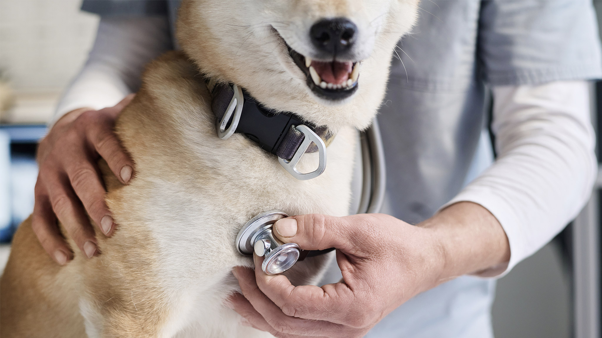
To take the HR using a stethoscope:
- If required, obtain assistance to restrain the patient.
- Place the stethoscope over the left side of the chest – the best access is usually over the rib cage, just behind the front left leg.
- Listen carefully and count the number of the thumps over 15 seconds then multiply by 4 to calculate beats per minute (bpm).
- Record this information.
To take the HR without a stethoscope:
- Place your hand over the left side of the animal’s chest. Depending on the side of the animal you can use your whole hand or just your middle and index fingers. Be careful not to use your thumb. You have a strong pulse at the tip of your thumb, which can make it difficult to detect the heart rate of the animal.
- Feel for the heartbeat.
- Once you have a good hand position, count the number of the thumps over 15 seconds then multiply by 4 to calculate bpm.
- Record the HR.
Pulse Rate
The pulse can be measured on any part of the body where a major artery runs close to the surface of the skin. Each pulsation corresponds with a contraction of the heart. If you do not have access to a stethoscope and have difficulty finding the heartbeat with your hands, you can take a pulse rate.
The two easiest places to find a pulse in most animals are at the femoral artery (1) and the dorsal pedal (2).
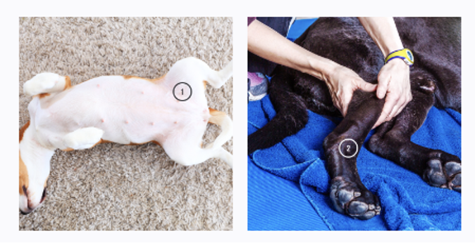
To find the femoral artery:
- Run your hand along the inside of the hind leg until you are almost to the point the leg joins with the body.
- You will feel a slight dip where the femoral artery is closest to the skin.
- The artery feels like a thin cord between two muscles.
A pulse is generally stronger the closer it is to the heart. So, the femoral artery is usually easier to locate than the dorsal pedal pulse. However, if you can’t access the femoral artery, you can try the dorsal pedal pulse.
To find the dorsal pedal pulse:
- Place two fingers (not your thumb) on the top (dorsal surface) of the hind foot or on the bottom (plantar) surface of the foot, just below the main pad. You need to apply a little more pressure to feel this pulse point.
- Once you locate one of the pulse points, use your fingertips (not your thumb) to press down gently and feel for a pulse. As with taking the heart rate, count the number of pulses over 15 seconds then multiply by 4 to calculate bpm.
HR outside the normal range
It is important to understand what a normal HR is for the particular species and breed of animal you are working with. The size and age of the animal may also affect the ‘normal range.’
- A higher than normal HR (tachycardia) could result from stress, a heart condition, anaphylaxis, blood loss or shock.
- A lower than normal HR (bradycardia) may indicate many things including a heart condition, congenital disorder, poisoning or hypothermia. Some medications also lower HR.

Respiratory rate (RR) is the number of breaths an animal takes in a minute. To check the patient’s RR:
- Watch the chest rise and fall or listen closely to the animal’s breathing.
- Count the number of breaths for a period of 15 seconds and multiply this number by 4.
You can assess the patient’s breathing at the same time as you take the RR during your distance assessment.
RR outside the normal range
Heat stroke, airway obstruction, fluid build-up in the lungs and some heart problems are common health issues that can cause a significant increase in RR.
Medication or anaesthetic overdose, apnea (temporary pauses in breathing, common during sleep) and some toxins and poisons will cause a significant decrease RR.

Mucous membranes (MM) are a type of tissue that line body cavities, such as the mouth. They typically contain a dense network of capillaries, and so are a good indicator of blood flow and of how much oxygen is circulating in the blood of an animal.
You can assess the MM of an animal by checking the colour of its gums.
- Moist and pink – normal and healthy
- Pale – may indicate shock or reduced blood flow
- Yellow, also known as jaundice – may indicate problems with liver function
- Blue – cyanotic (lack of oxygen) – may indicate poor breathing quality or poor circulation
- Bright red – may indicate poisoning or heatstroke.
Review the following video, for a demonstration of how to check the MM of a dog. The process is essentially the same for all mammals.
Watch
Checking your pet's mucous membrane colour
This video, featuring Dr Gemma Spinoglio demonstrates how to check your pet's mucous membrane colour at home.
Expected Duration: 3:01 minutes
Questions
Pre Watch Question: What are the specific health conditions that Dr Spinoglio mentions she looks for when checking mucous membranes? Make a note when you hear them.
Capillary refill time
Blood flows continually around the body. The speed at which it flows can be measured using capillary refill time (CRT).
To measure CRT
- Gently compress small blood vessels (capillaries) near the surface of the skin to stop the blood from flowing through.
- When you release the pressure, blood will start to flow again.
- The time taken between lifting the pressure and blood refilling the capillary is the CRT.
One of the most common places to check CRT is on the gum of an animal, as long as it is safe to put your finger in the animal’s mouth. CRT should only take 1-2 seconds in a healthy animal. Longer than this may indicate dehydration, low blood pressure, circulatory problems or blood loss.
To check the CRT:
- Lift the animal’s lip.
- Apply a small amount of pressure with the tip of one finger to the side of the gum. As you press down, the blood leaves the area and makes the gum appear pale - blanches.
- Remove your finger and observe how long it takes for the gum to return to a pink colour.
You can assess CRT at the same time as you check the MM.

The most accurate method of taking a patient’s temperature is with a digital rectal thermometer.
Note: Taking temperature with a rectal thermometer is the most invasive technique for checking vital signs. The discomfort will increase the animal’s HR and RR. So, it should be the last vital sign you check during the health assessment.
Taking an animal’s temperature
Many animals will require some form of restraint when having their temperature taken. As always, use the minimal amount of restraint required for that animal. Ideally, one person should hold the animal’s head while a second person should take the animal’s temperature. Always lubricate the thermometer to make the procedure more comfortable for the animal.
To take an animal’s temperature using a rectal thermometer:
- Place a clean probe cover on the thermometer and lubricate the bulb.
- Lift the tail and gently insert the thermometer into the rectum, slightly rotating it as you go. Do not force entry. Gently push the thermometer against the wall of the rectum – you are taking the temperature of the animal, not its faeces!
- Keep the thermometer against the wall of the rectum for a minimum of 60 seconds (or until you hear the beep sound on some thermometers). For large animals, ensure the thermometer stays in contact with the wall of the rectum for a correct reading. Most digital thermometers will beep when you turn them on, and beep again when it’s ready to read – after maintaining a constant temperature for several seconds.
- Gently remove the thermometer. Immediately read the temperature and record the finding.
- Dispose of the probe cover appropriately, wipe away any faeces and wash and sterilise the thermometer before using it again.
Temperatures outside the normal range
When an animal’s temperature is significantly lower than the normal range, the animal is said to be hypothermic. Hypothermia can be caused by prolonged exposure to cold conditions, some diseases, blood loss and even anaesthetic.
Symptoms of hypothermia include:
- In mild cases:
- hivering
- stiff or clumsy movement
- pale gums
- confusion.
- In severe cases:
- no shivering
- rapid decrease in body temperature
- slow breathing
- fixed and dilated pupils
- collapse or lack of responsiveness.
When temperatures are higher than normal, the animal is hyperthermic. A high temperature is a common indication of infection. Hyperthermia can also be caused by extreme environmental heat, such as being locked in a car on a hot day or by excessive activity on a hot day.
Hyperthermia can cause serious damage to the body and quickly lead to death if not treated.
Early symptoms of hyperthermia include:
- Panting
- Drooling
- Bright red MM.
Symptoms of severe hyperthermia include:
- Seizures
- Spontaneous bleeding
- Extreme lethargy
- Collapse
- Coma and death.
Summary
You now know how to check:
- Heart rate
- Pulse rate
- Respiratory rate
- Mucous membrane
- Temperature
For each of the vital signs you can also identify what indicates good health and what indicates that something might be wrong. This is a really important practical skill to master. Tau kē!
Activity
Review what we have covered in this topic by completing the following activity. For some of the questions more than one answer will apply. You will need to refer back to some of the details in the learning content for specific answers.