Nau mai, hoki mai, welcome back!
This week, we will continue our journey through the digestive system before flowing into the system that influences almost every part of the human body, the endocrine system. We will also pick up where we left off with the nervous system and cardiovascular systems.
By the end of this week, you will have explored the anatomy of four more body systems and discovered several more common disorders and treatments for these body systems to your rapidly growing kete (basket) of knowledge.
Kia ora, welcome to this session of Patient Care 2, where we continue our learning on the anatomy of body systems and how they relate to the treatment of health disorders.
In our last session, we explored the nervous system and the digestive system. Kia haere tonu tātou, let’s continue now with learning more about the digestive system.
The digestive system - part 2

As we did last week, you may wish to take a moment now to refresh your memory on what we’ve covered so far.
Digestive system review
In our last session, you watched a video that gave you an overview of the structures and functions of the digestive system. You might like to watch this again to prep yourself for the following activity: GCSE Biology – Digestive System #18.
In this activity, challenge yourself to have a go at remembering the names, locations and functions of the parts of the digestive system. We don’t expect you to remember them all. This is NOT a test! It is just one way to help you recall and retain this important information.
How did you go? We hope this has helped you feel more confident in locating the different parts of the digestive system and how they all function together.
You might like to keep the completed diagram handy for future reference. Click here to download and save it: Digestive System Review - Labelled Diagram.
Stomach

The stomach is in the upper left of the abdomen, positioned between the oesophagus and the small intestine. As mentioned previously, the lower oesophageal sphincter, located between the lower oesophagus and the upper stomach, acts as a barrier, contracting to prevent the reflux of acidic stomach contents into the oesophagus.
The outer walls of the stomach are composed of muscles that help the stomach expand during eating and contract to mix and churn digested food into a substance called chyme.
The inner lining of the stomach is called the mucosa. The mucosa produces pepsin, an enzyme that breaks down protein. It also produces hydrochloric acid (HCL) to aid the action of pepsin and kill bacteria.
A layer of mucus produced by the mucosa protects the stomach lining from the corrosive effects of stomach acid.
There is a small amount of absorption from the stomach into the bloodstream of water, salts, alcohol and some medicines.
The muscular contractions of the stomach wall, known as peristalsis, move the chyme toward the small intestine.
Between the bottom of the stomach and the small intestine is a sphincter known as the pyloric sphincter, whose role is to regulate the release of chyme into the small intestine. It also helps prevent the backflow of small intestine contents into the stomach.
Stomach anatomy
Can you spot the oesophagus, the stomach’s outer muscle layer, the inner lining (mucosa), and the pyloric sphincter?

Small intestine
The small intestine is a hollow tube about 3-5 metres long (in an adult) coiled within the abdominal cavity. The walls are made up of several layers.
The muscular layer contracts to mix and move chyme through the small intestine towards the large intestine.
The inner lining has finger-like projections known as villi, which are made of tiny folds called microvilli. These projections increase the surface area of the small intestine, allowing faster absorption of nutrients into the bloodstream.
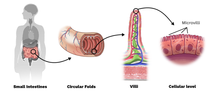
As well as villi and microvilli, the inner lining also has circular folds called plicae circulares which also increase the surface area for absorption of nutrients.
Main segments
The small intestine has three main segments that form a continuous tube. Click on the (+) symbol to expand the labels below and read more about each segment and its functions.
- The first segment immediately follows the stomach.
- Receives chyme from the stomach.
- Receives bile from the liver and pancreatic juices from the pancreas.
- Neutralises acidic chyme.
Fun fact – Duodenum means ‘12 fingers’ in Latin (which is roughly its length!).

- The second segment follows the duodenum.
- Absorbs nutrients such as amino acids, sugars, and other small molecules.
Fun (or not so fun fact) – Jejunum comes from the Latin word jejunus, which means ‘fasting’. Apparently, this is because this segment of the small intestine is often found to be empty after death.

- The last segment connects to the large intestine (cecum).
- Continues absorbing nutrients, particularly vitamin B12, bile salts, and any remaining nutrients not absorbed in the jejunum.
Fun fact – In Greek, Ileum means to roll or twist.

Pancreas
The pancreas plays an important role in both the digestive and endocrine systems.
Here, we focus on its digestive function, and later in this week’s Anatomy and Physiology session, we will reintroduce ourselves as we explore its additional roles in the endocrine system.
The pancreas is located below and behind the stomach in the abdominal cavity. The head and neck of the pancreas lie slightly to the right of the middle of the body while the tail extends towards the left side.

It is mainly made up of exocrine glands that secrete enzymes to aid food digestion in the small intestine. The main enzymes produced are lipases, peptidases and amylases to break down fats, proteins and carbohydrates. They are transported from the pancreas via the pancreatic duct to the duodenum.
Liver and gallbladder

The liver is located in the upper right area of the abdomen below the diaphragm.
The liver produces bile, a greenish-yellow fluid that contains bile salts, cholesterol, bilirubin, and other substances. Bile aids in emulsifying fats, breaking them down into smaller droplets, which aids their digestion by pancreatic enzymes.
The gall bladder is located beneath the liver, tucked within a small hollow on the liver's surface. Bile is stored and concentrated in the gallbladder before being released into the small intestine.
You can see in the diagram that the bile duct goes through the pancreas and joins with the pancreatic duct just before entering the duodenum.
Large intestine

The large intestine, approximately 1.5 metres in length in adults, is a hollow tube with a wider diameter and shorter length compared to the small intestine. Positioned in the right lower abdomen, the ileocecal valve marks the boundary between the last section of the small intestine and the first part of the large intestine. The valve acts as a regulator, overseeing the movement of digested material from the small to the large intestine while preventing backflow.
Main segments
Here's an overview of the large intestine – click the labels or the (+) icon to expand each segment.
It receives and temporarily stores undigested material from the small intestine. Think of it like a short-term storage container.
Fun fact – cecum derives from the Latin intestinum caecum, which means ‘blind gut’ or ‘cul de sac’. When you see the image of where the cecum is, this makes sense!
Its exact function in humans is not fully understood. It is believed to have immune-related functions.
The colon is the largest part of the large intestine and is divided into segments. These are the:
- ascending colon: travels up the right side of the abdominal cavity.
- transverse colon: travels across the abdominal cavity.
- descending colon: travels down the left side of the abdominal cavity.
- sigmoid colon: s-shaped part of the colon that lies in the pelvic cavity.
Functions
As you can see, a bit is going on in the colon. Let’s break down the functions of this impressive organ:
- The colon absorbs most residual water, electrolytes, and vitamins from the chyme into blood bloodstream.
- This transforms the chyme into the solid formation known as faeces.
- Faeces is composed of undigested food residues, unabsorbed digested substances, bacteria, old cells from the GI mucosa, inorganic salts, and enough water to let it pass smoothly out of the body.
- The colon then temporarily stores the faeces.
- Peristaltic movements of the intestine wall move the faeces toward the rectum.
Rectum
The rectum is a muscular tube that connects the sigmoid colon to the anus. The rectum's muscular walls help the compaction and storage of faeces. The rectum sends signals to the brain if faeces are to be evacuated and holds stool until evacuation can happen.
Anus
The anus consists of pelvic floor muscles and two anal sphincters:
- Inner/internal sphincter: under involuntary control.
- Outer/external sphincter: under voluntary control.
Functions
The sphincters keep the anus closed as stool collects in the rectum. Eventually, the pressure on the rectum wall causes the internal anal sphincter to relax. Conscious control over the external anal sphincter allows stool to pass out of the body through the anus at an appropriate time.

Disorders and treatments
Throughout the last few weeks, you will have gathered quite a bit of knowledge about digestive system disorders and their treatments. Now, use this knowledge as a foundation to research and seek answers about other common digestive disorders.
Journal post
Digestive system: Disorders and treatments
This activity has two purposes. You will learn new information through self-discovery AND create your own learning resource.
- Use reliable resources to research and complete the Documentation tool activity.
- Download and save the completed activity.
- Create a journal post titled ‘Digestive System: Disorders and Treatments.’
- Congratulate yourself and upload the completed activity Word document to the journal post.
- As your tutor will review your post, make sure to check back later for feedback.
The endocrine system

The endocrine (hormone) system is the system responsible for internal communication between organs and tissue. It does this through the release and production of hormones. These hormones act as chemical messenger pigeons, sending orders to the tissues and organs to decrease or increase activity.
Watch: GCSE Biology – Endocrine System & Hormones #59 (5:03 minutes)
Watch this video for a brief overview of the anatomy and physiology of the endocrine system. Āta whakairo, think carefully and take notes as you watch and apply this knowledge to an activity following.
Journal post
The endocrine system
- Create a new journal post titled ‘The Endocrine System’.
- Answer the following questions using the information from this video and your own research.
- As your tutor will review your mahi, publish your post to ‘All course users’.
- And of course, save the permalink to your Index of Journal Posts. You want to keep track of all your hard work!
Questions

- What is an endocrine gland?
- What is the main function of the endocrine system?
- How does the endocrine system differ from the nervous system? Create a list comparing the two systems.
- What is the main function of the endocrine system?
- What is a hormone?
- What gland is known as the master gland, and where is it located?
- What gland releases adrenaline/ epinephrine, and where is it located?
- What hormone does the pancreas produce?
- What endocrine glands are part of the male reproductive system, and which are part of the female reproductive system?
- Name three disorders of the endocrine system.
By now, you will be able to identify the glands throughout the body that make up the endocrine system, describe their functions, and list the differences and similarities between the nervous and endocrine systems.
Before you finish this week’s learning, apply this knowledge to an endocrine disorder of your choice. This activity will allow you to hone in on how the endocrine system functions in a specific way.
Self-directed learning activity
- Create a new journal post, titled ‘SDL – Endocrine Disorder’.
- Choose one of the three endocrine disorders you named in the last learning activity, for example, hypothyroidism, Graves’ disease, or Addison’s disease.
- For that disorder, write a brief information report covering the following points:
- What part/s of the endocrine are affected by this disorder, and how are they affected?
- What are the signs and symptoms of this disorder?
- What are the causes of this disorder?
- What pharmaceutical treatments are used to treat or manage this disorder?
- Publish your answers to ‘All course users’. Remember to check your work for tutor feedback later in the week.
- Save the permalink to the Index of Journal Posts.
And just like that, this session of Patient Care is complete. Ka mutu pea, you’re doing awesome!

Malo e lelei, welcome to this session of Anatomy and Physiology, where we continue to explore disorders of the cardiovascular system, build on our knowledge of the nervous system and apply this knowledge to treat different health disorders.
Cardiovascular (CV) disorders and treatment
So far, we have looked at the following CV disorders and their treatment:
- Hypertension
- Dyslipidaemia and hypercholesterolemia
- Atherosclerosis
- Thrombosis and embolism
- Ischemic Heart Disease (IHD)
- Angina pectoris
- Myocardial Infarction (MI)
- Congestive Heart Failure (CHF).
Kia haere tonu tātou, let’s continue to look at disorders and treatment (as well as introduce new acronyms for your glossary!).
Arrhythmia
Arrhythmias are a disorder that results from irregular generation or conduction of electrical signals in the heart. These signals coordinate the heart muscle's rhythmic contractions, ensuring efficient blood circulation for oxygen and nutrient delivery to the body's tissues, including the heart. However, arrhythmias can cause the heart to beat too fast, too slow, or irregularly. This disrupts normal blood supply and flow, leading to inadequate oxygen and nutrient delivery to the body and heart muscle.
Causes
Various factors, including heart disease, high blood pressure, diabetes, smoking, excessive alcohol consumption, and aging can cause arrhythmias. Some arrhythmias may not cause noticeable symptoms, while others can lead to dizziness, fainting, chest pain, or life-threatening complications.
Types of arrythmias
There are various types of arrhythmias, including:
- Tachycardia: Abnormally fast heart rhythm.
- Bradycardia: Abnormally slow heart rhythm.
- Atrial Fibrillation (AF): Irregular and often rapid heartbeat.
- Atrial Flutter: Rapid, regular atrial contractions.
- Ventricular Tachycardia: Fast, regular beating of the heart's lower chambers.
- Ventricular Fibrillation: Chaotic, rapid beating of the heart's lower chambers, a medical emergency.
Atrial Fibrillation (Manawa tukituki)
Atrial Fibrillation (AF) is a common form of arrhythmia that can cause poor blood flow.
Watch: What is Atrial Fibrillation? (2:12 minutes)
As you watch this video, note the symptoms some people may experience. Check your answers in the activity that follows.
Abnormal impulses
In this illustration, you can see the abnormal impulses in the atria.
Under the drawing of the heart, we can see the difference between a normal heart rhythm recorded in an electrocardiogram (ECG) and one that shows the disorganised electrical conduction in a person with AF.

Treatment of AF
The pharmaceutical treatment for AF may include the following medicines (some of which you may already be familiar with).
Antiarrhythmics
Antiarrhythmics are a group of medicines designed to prevent or treat abnormal heart rhythms. They are divided into four different classes.
Expand each label for more information.
- Example: Flecainide.
- Action: Rhythm control - to control an irregular heart rhythm.
- Example: Metoprolol, Bisoprolol, Carvedilol.
- Action: Rate control - to slow a fast heart rate.
- Example: Amiodarone.
- Action: Rhythm control - to control an irregular or fast heart rate by slowing the electrical signals through the heart.
- Example: Verapamil, Diltiazem.
- Action: Rate control - to slow a fast heart rate.
How well can you recall the action for each class of medicine? Complete this activity to aid your retention.
Anticoagulants
According to the Stroke Foundation of NZ (n.d), people with AF are five times more likely to have a stroke than those who do not. This is due to ineffective atrial contractions, causing blood stagnation in the heart chambers. Stagnant blood can lead to clot formation, with the potential for these clots to be pumped out of the heart, where they travel through the bloodstream to the brain and other parts of the body.
Anticoagulants are used to reduce the likelihood of blood clots forming.
- Example: Warfarin.
- Action: Interferes with the production of vitamin K-dependent clotting factors in the liver to reduce clots forming.
Other anticoagulants that may be used instead of warfarin are Dabigatran and Rivaroxaban. These act directly on key components of the blood clotting process, providing anticoagulant effects without affecting vitamin K metabolism.
Other medications used in treating AF
If other medications are not effective in treating AF, cardiac glycoside medications may be used and also considered in patients with congestive heart failure.
- Example: Digoxin.
- Action: Strengthens the force of contraction of the heart muscle, which leads to more blood being pumped out of the heart with each heartbeat.
Journal post
Treatments for Atrial Fibrillation
In this activity, you will step back into the shoes of a pharmacy technician and conduct your own rangahau (research) to support and answer your patients with confidence.
- Complete the following Documentation tool activity.
- Download and save the completed activity.
- Create a journal post titled ‘Treatments for Atrial Fibrillation’.
- Upload the completed activity/ Word document to the post and publish it to ‘All course users’.
- Return to the post in the next week for tutor feedback.
Anaemia
Anaemia is a disorder of the blood. Try answering these questions to help you learn more about this disorder. If you don’t get the correct answer, no sweat! Instead, note, draw or use your preferred learning method to ensure you understand the correct answer.
Types of anaemia
Anaemias can be categorised in different ways based on different criteria. We will look at them under the following four types.
Expand each label for more information.
- Occurs when the body does not have enough iron to produce haemoglobin.
- Without the iron in the haemoglobin molecule, oxygen cannot attach to haemoglobin in red blood cells, and as a result, the crucial transport of oxygen throughout the body is compromised.
- Occurs when the body cannot make enough red blood cells because it cannot absorb enough vitamin B12 from food.
- Vitamin B12 is required to form healthy red blood cells in the bone marrow and produce DNA, which is essential for properly functioning and maintaining the nervous system.
- When there are not enough healthy red blood cells circulating in the blood, less oxygen is transported to the body's tissues.
- Occurs when the body cannot make enough red blood cells, white blood cells and platelets.
- This results in a decreased ability of the blood to carry oxygen, weakened immune function, and impaired blood clotting.
- Occurs when the body destroys red blood cells and cannot replace them quickly enough.
- This results in decreased red blood cells circulating in the bloodstream and an increased workload on the bone marrow to produce more red blood cells.
- The accelerated destruction of red blood cells can contribute to the release of bilirubin, causing the characteristic yellowish discolouration associated with jaundice.
Treatment of anaemia
Treatment for anaemia will depend on the underlying cause and the specific type of anaemia the patient has been diagnosed with. One medicine used in treatment is iron tablets.
Watch: Iron Tablets | How To Take Iron Tablets […] (3:25 minutes)
Iron tablets are a fairly common supplement used for treating anaemia. However, do you know how to take them, their common side effects, and how to reduce these? If not, join our friend Abraham The Pharmacist, who covers this and more.
As you know, a fantastic way to learn something is by doing. Apply your knowledge of this topic to a real-life scenario!
Journal post
Anaemia
It’s time to again step into a pharmacy assistant's shoes and support a patient in a low-risk way that simulates the real-world. This activity is a perfect way to have the luxury of researching the correct information and considering how you would present this to a patient.
- Complete the Documentation tool activity below.
- Download and save the completed activity.
- Create a journal post titled ‘Anaemia’.
- Upload the completed activity/ Word document to the post and publish it to ‘All course users’.
- Return to the post in the next week for tutor feedback.
Mahi nui! Great work! That completes our exploration of the cardiovascular system for Anatomy and Physiology. Now, to the nervous system…
Nervous System Part 1

In Patient Care 2, we discussed how the nervous system is organised. We looked at both the central and peripheral nervous system and their divisions. We noted that the function of the nervous system is to regulate and coordinate body functions such as:
- Receiving, processing and responding to information
- Maintenance of homeostasis
- Body movement
- Learning and memory
- Survival responses.
We will build on that learning and continue to learn about the anatomy, physiology, disorders and treatments related to this body system.
Neurons

You can see in this graphic that neurons are organised in a chain. The axon terminal of one neuron is connected to the dendrites of the next by the synaptic cleft.
As mentioned previously, neurons are the basic building blocks of the nervous system. They are specialised cells that transmit electrochemical signals in one direction, from one end of their structure (the dendrites) to the other (the axon terminals).
Neurons are organised in long chains where the axon terminals of one neuron lie very close to the dendrites of the next. The small space between one neuron and the next is known as the synapse or synaptic cleft and is sometimes referred to as the synaptic gap. This chain-like organisation allows the transmission of information from one neuron to the next.
Watch: 2-Minutes Neuroscience: The Neuron (1:47 minutes)
For a simple explanation of the neuron, its structure, functions and how it transmits signals, mātakitaki (watch) this video. Pay attention to the names of the neuron anatomy, as there will be a short quiz that follows.
Ready? Karawhiua, give it your best shot! Remember, this is a learning opportunity, so if you’re unsure of the answers, watch the video again or write down some questions to take to your tutor or pharmacy colleagues.
Electrochemical transmission of signals

In the video, you will have seen the electrochemical transmission of signals in neurons. Signals entering a neuron via its receptors on its dendrites arrive as chemical neurotransmitters, which then travel to the neuron's cell body.
In the cell body, the neurotransmitter is interpreted, and if the signal is strong enough, an electrical impulse is generated. This impulse then travels along the axon to the axon terminals, which triggers the release of chemical neurotransmitters into the synapse.
The neurotransmitters travel across the synaptic cleft to bind with neighbouring neuron’s receptors on its dendrites. Due to the small size of the synaptic cleft (in the range of billionths of a meter), the transmission of signals between neurons is exceptionally fast!
Examples of neurotransmitters include:
- Serotonin
- Gamma-aminobutyric acid (GABA)
- Dopamine
- Noradrenaline
- Adrenaline
- Acetylcholine.
A nerve fibre is part of a neuron. It may be either:
- An axon = The long, thin part of the neuron that transmits signals from away from the cell body.
- Dendrites = The shorter, branched extensions of the neuron that receive signals from other neurons and carry them to the cell body.
A nerve is a bundle of fibres comprised of individual neurons, associated connective tissue, and blood vessels. Nerves are part of the peripheral nervous system, the network of nerves extending throughout the body beyond the brain and spinal cord.
Self-directed learning activity
As we only briefly touched on neurotransmitters in this session, it’s up to you to take your learning deeper.
- Choose one of the neurotransmitters listed in this session and determine its role in the human body.
- Summarise and share the findings in the forum: SDL: Neurotransmitters.
- Include references for any resources you used for your research.
- You may also choose to share links to any YouTube videos, articles or other educational sources that you find interesting.
You have now completed this week’s learning for Anatomy and Physiology. Ka mau te wehi, awesome. Our next session will focus on nervous system disorders and treatment.