Afio mai, welcome! The journey continues through the human body, with stop-offs to check out three more body systems and a drive through the pain pathway - an exploration of the nervous system. We will look at how humans perceive and respond to pain as well as common disorders, such as depression, anxiety, epilepsy, Parkinson’s disease and some of their treatments.
Caution
As we journey through this content, some people may find some of the disorders or content we cover confronting or triggering. If this happens, we encourage you to take the content slowly and at your own pace. Your tutor is always available to chat, and if you feel that you need additional support, you can book a session with a counsellor at www.acscounselling.com.au/book-appointment.
This week in Patient Care 2, we will explore the respiratory, reproductive and urinary systems.
The respiratory system
The function of the respiratory system is to work with the cardiovascular system to deliver oxygen to the body and remove carbon dioxide. This process is called ‘gas exchange’. Oxygen is essential for the body to perform its various functions and maintain homeostasis. Carbon dioxide is a waste product; if too much of it builds up in the body, it can lead to serious and life-threatening conditions.
While the major organs of the respiratory system function to provide oxygen and expel carbon dioxide, other important components include:
- the sinuses, which humidify and filter air in the nasal cavity.
- the mouth which can be an alternative entry point for air, particularly during activities like breathing through the mouth. It also plays a role in speech production.
- the diaphragm, which contracts to draw air into the lungs and relaxes to push air out.
Respiratory system organs
The major organs of the respiratory system are divided into two sections (again, creatively named): the upper respiratory tract and the lower respiratory tract.
Click on each label or the (+) symbol to read about the body parts that comprise each tract.
- Nose: The entry point for air into the respiratory system.
- Pharynx: The throat, where the pathways for air and food cross.
- Larynx: The voice box, containing vocal cords and aiding in sound production.
- Trachea: A tubular structure that carries air from the pharynx to the lungs.
- Bronchi: Two branches of the trachea that lead to the left and right lungs. Note: bronchi is the plural for bronchus (one branch).
- Lungs: Paired organs responsible for gas exchange, where oxygen is taken in and carbon dioxide is expelled.
- Bronchioles: Smaller airways branching within the lungs.
- Alveoli: Tiny air sacs within the lungs where gas exchange occurs between air and blood vessels.
Let's take a closer look at air passage through the respiratory system while noting each structure and its functions.
Upper respiratory tract
Nose
Air from the environment is breathed in via the nose. The nose filters this air by trapping unwanted particles, preventing them from entering the rest of the body.
Particles are trapped by the cilia, tiny hairs inside the nose and in the sticky mucous that is produced by the nasal lining (nasal mucosa). The nose also warms and moistens the incoming air before it passes through to the pharynx.
Pharynx
The filtered, warmed, and moistened air then travels to the pharynx (throat). Funnel-shaped and approximately 13cm long, the pharynx is composed of skeletal muscle and lined with a mucous-producing membrane.
The pharynx is divided into three regions:
- Nasopharynx
- Oropharynx
- Laryngopharynx.
The pharynx is also part of the digestive system as it receives chewed food from the mouth on its way to the oesophagus, which lies behind the larynx and trachea.
Larynx
From the pharynx, the air passes into the larynx or voice box. It is formed by nine cartilage rings that are connected to each other by muscles and ligaments. The main function of the larynx is to produce sounds and speech.
As you can see in this image, at the top of the larynx is a flap of cartilage called the epiglottis. When food is swallowed, the larynx is pulled upward, and the epiglottis tips, forming a lid over the larynx’s opening.
This prevents food from entering the larynx and directs it to the oesophagus for its passage to the stomach. If anything other than air enters, the cough reflex is triggered to expel the substance and prevent it from continuing into the respiratory system.
Ka pai, good work. We’ve now covered the upper respiratory tract, so let’s move down into the lower respiratory tract.
Lower respiratory tract
Trachea
From the larynx, air passes into the trachea or windpipe. The trachea is made up of fibrous and elastic tissues and smooth muscle with about twenty rings of cartilage, which help keep the trachea open. It is lined with goblet cells that secrete mucous and other cells with very small hair-like fringes called cilia.
The mucous traps tiny particles of debris, and the cilia move the mucous up and out of the respiratory tract, keeping the lungs and airways clear. The trachea then divides into two branches called the main bronchi.
Bronchi
The two branches of the bronchi are called the left main bronchus and the right main bronchus. They enter the lung tissue to carry the air to the lungs. Each main bronchus divides further into secondary and tertiary bronchi. These many branches ensure air is passed to all parts of the lung.
Lungs
The left and right lungs are the main organs of the respiratory system. They lay in the chest surrounded and protected by ribcage. The left lung is smaller and has two lobes to accommodate the heart, while the right lung is larger and has three lobes.
Bronchioles
Deep inside the lungs, the branching bronchi form into bronchioles. Air travels through the bronchioles and enters the alveoli.
Alveoli
The inhaled air is now in the alveoli, tiny air sacks with very thin walls. The oxygen molecules in the air pass through the alveoli walls and enter the bloodstream, while at the same time, carbon dioxide passes from the bloodstream into the alveoli. This process is called gas exchange.
The oxygen is now in the cardiovascular system and travels in the bloodstream to be delivered to body tissues. Meanwhile, the carbon dioxide travels from the alveoli to the bronchioles and all the way through the respiratory system to the mouth and nose, where it is exhaled into the environment.
Ka pēhea ōu whakaaro e pā ana ki tēnā? How do you feel about that? You’ve now wrapped up your learning about the anatomy of the respiratory system and learned how it impacts and connects to some of the other body systems (such as the digestive system).
What do you know?
Now that you’ve completed this section, connect this content to everything you’ve learned throughout the programme on the respiratory system. Using your knowledge of the respiratory system, the different respiratory disorders and their treatments, take this multichoice quiz to check what you know. Watch out! One of the questions may have more than one answer!
The reproductive system
The primary function of the male and female reproductive systems is to ensure the continuation of the species through producing reproductive cells.
Female reproductive system
The female reproductive system has two functions:
- Produce egg cells for fertilisation.
- Protect and nourish the foetus until birth.
We will focus on the following main structures that make up this system. Click on each label or the (+) symbol for an overview of the anatomy and its function.
Small glands are attached to each side of the uterus by a thin, fibrous ovarian ligament. They are responsible for storing and nurturing immature egg cells into mature eggs. Each month, one of them releases a mature egg into its fallopian tube.
The ovaries also produce two female sex hormones - oestrogen and progesterone, which are involved in regulating the menstrual cycle.
These tubes connect the ovaries to the uterus. The fringed fimbriae capture the ovum (egg cell) when it is released from the ovary and draw it into the fallopian tube. The ovum travels along the fallopian tube, where it may be fertilised by the sperm.
This is a hollow, pear-shaped organ with a thick muscular wall. It lies in the middle of the pelvis, between the bladder and the rectum. The endometrium is the lining of the uterus. Roughly every 28 days, it thickens in preparation for a potential pregnancy and then sheds during menstruation if pregnancy does not occur.
If a pregnancy occurs, the fertilised ovum implants in the endometrium, and the pregnancy begins. The uterus protects the foetus while it grows and develops. It contracts during childbirth to deliver the baby.
The cervix joins the uterus to the vagina. It is important to allow fluids to pass between the uterus and vagina. For example, sperm enters the uterus through the vagina during sexual intercourse, and menstrual fluid flows out during menstruation.
This is a muscular, narrow canal that extends from the vaginal opening to the cervix. The inner wall of the vagina has folds of soft elastic mucous membranes that allow the vagina to expand during sexual intercourse or childbirth.
Male reproductive system
The main function of the male reproductive system is to:
- Produce sperm to fertilise the eggs that the female produces.
It is made up of the following structures: Click on each label or the (+) symbol for an overview of the anatomy and its function.
The penis has three cylindrical spaces of erectile tissue: the two larger ones, called the corpora cavernosa, sit side-by-side, while the third, the corpus spongiosum, surrounds most of the urethra. When these spaces fill with blood, the penis becomes erect.
The scrotum is the thick-skinned sac that surrounds and protects the testes. The scrotum also acts as a climate-control system for the testes, as they need to be slightly cooler than body temperature for normal sperm development.
The scrotum can hang farther from the body to cool or contract to pull the testes closer to the body for warmth or protection.
The testes (testis - singular) are oval and produce sperm (sex cells) and testosterone. The testes are also known as testicles.
This is a long, coiled tube that collects sperm from the testes and stores it for maturation, then transports it into the vas deferens. One epididymis lies against each testis.
This is a tube, one from each testis, that transports sperm from the epididymis to the back of the prostate and joins with one of the two seminal vesicles.
The urethra is a tube that runs through the shaft of the penis, providing an outlet for both urine and semen.
This gland lies just under the bladder and surrounds the urethra. The fluid produced by the prostate helps nourish and transport sperm during ejaculation.
Pea-sized structures on the sides of the urethra, just below the prostate. They create a clear, slippery fluid that empties directly into the urethra. This fluid lubricates the urethra and neutralises any acids that may remain from urine.
These are located above the prostate and join with the vas deferens to form the ejaculatory ducts, which travel through the prostate. The seminal vesicles produce fluid that nourishes the sperm. This fluid provides most of the volume of semen.
You will learn more about the reproductive system and common disorders and treatments in an upcoming week of Anatomy and Physiology.
The urinary system
The urinary system filters out excess fluid and other substances from the bloodstream as urine. Urine is produced by the kidneys, collected in the bladder, and excreted through the urethra.
The urinary system includes the following organs. Click on the (+) icon for an overview of the anatomy and function of each.
The kidneys are a pair of bean-shaped organs located at the rear wall of the abdominal cavity just above the waistline and protected by the ribcage. Within the kidney are structures called renal pyramids. These pyramids consist of numerous nephrons and microscopic filters that are crucial in blood filtration.
Nephrons efficiently separate waste products and excess fluids from the blood, forming urine as the end product, which is drained from the kidney into the ureters. The nephrons regulate electrolyte balance, maintain fluid levels, and control blood pressure through this filtering process.
The kidneys also play a pivotal role in the endocrine system by producing hormones such as erythropoietin, which stimulates red blood cell production and renin, which is essential for blood pressure regulation.
This is an internal view of the kidney. See if you can identify the structures in bold and visualise their role in this system.
The ureters are two tubes that drain urine from the kidneys to the bladder. Muscles in the walls of the ureters send the urine in small spurts into the bladder.
The bladder is an expandable storage sac that receives urine from the kidney via the ureters. When full, sensory nerves in the bladder wall are stimulated to relax the internal urethral sphincter at the base of the bladder. This involuntary sphincter keeps the urethra closed when urine is not being passed.
The urethra is a thin-walled, muscular tube that carries urine by peristalsis* from the bladder to the outside of the body. It is shorter in a woman than in a man.
The external urethral sphincter is located where the urethra passes through the pelvic floor muscles. The sphincter is under voluntary control and contracts to prevent urine from exiting the body until it is an appropriate time to let the sphincter relax and allow the bladder to empty.
*Do you know what peristalsis means? If not, take a moment now to look up this word and add it to your glossary.
Locating the urinary system
This is the location of the urinary system in the body.
Identify each organ and take note of how this system links to the cardiovascular system.
That’s it for the urinary system this week! You will continue your exploration of this system in an upcoming week of Anatomy and Physiology.
Self-directed learning activities
To consolidate your learning of the respiratory, reproductive and urinary systems, there are three activities to complete (and an optional ‘Level-Up’ activity if you want to challenge yourself!).
Activity 1
Watch: How Do Your Lungs Work (6:59 minutes)
Pull together what you’ve learned this week about the anatomy of the respiratory system and see it in action. Transport yourself into the human body and watch how the lungs work in this cool 3D video. Use your learning method of choice to capture and connect new information to your existing knowledge. This may include writing keywords, drawing diagrams, or exploring related videos.
Activity 2
Watch: The Human Reproductive System (11:13 minutes)
Watch this video for a simple inquiry into the human reproductive system as Professor Dave asks the age-old question – how do humans make more humans? As you watch, take notes of information that is new to you and then answer the following questions:
Activity 3
Watch: The Structure and Function of the Nephron – Made Easy – Kidney Function (4:38 minutes)
To enhance your understanding of the nephron, watch this video. The knowledge and insights you acquire will be foundational in building your understanding of the urinary system, which we will delve into further during your upcoming Anatomy and Physiology course.
As you watch, add the words below to your (ever-expanding) glossary and include their meanings or a description. You may like to research these words to ensure you understand what they mean.
Important
You will need to access these words later for revision. Ensure you are saving your glossary in an easy-to-find location, e.g. in a folder on your desktop or in OneNote, Trello, etc.
Glossary terms
- Filtration
- Reabsorption
- Secretion
- Elimination
- Proximal tubule
- Distal tubule
- Collecting duct
- Renal pelvis
Level-Up Activity (Optional)
Without referring to any resources, challenge yourself to draw these three body systems (you can choose either the male or female reproductive system, or both!) without referring to your notes. You can do this on a scrap of paper or use an online template to get you started. Give yourself a point for each organ that you locate and label correctly.
You have now completed Anatomy and Physiology for this week – ka mau te wehi, awesome! Don’t forget to complete your SDL activities, as these will help you consolidate your learning and prepare you for future learning.

Haere mai, welcome to this week’s session on Anatomy and Physiology, where we will look at nervous system disorders and treatments.
Nervous system disorders and treatment
By now, you will have a good understanding of the anatomy and physiology of the nervous system. This week, we will build on this understanding to learn more about this system’s disorders and treatments.
Pain and treatment
The pain pathway
Pain is perceived, interpreted and responded to by the nervous system. Select each label or the (+) icon to take a closer look at the ‘pain pathway’.
Incoming pain signals travel along the nervous system's sensory (afferent) pathway.
- Specialised “receptors” in the skin and internal organs called nociceptors detect noxious (harmful or damaging) stimuli. Stimuli are factors such as temperature, tissue damage or pressure.
- Nociceptors generate signals that travel along sensory peripheral nerves to the spinal cord, and from there, they travel up to the brain's thalamus and somatosensory cortex.
- After the pain signals reach the brain, they undergo interpretation in the cerebral cortex.
- The brain analyses the sensory information, considering factors such as intensity, location, and emotional context.
- Drawing from past experiences and memories, the brain assigns meaning to the pain stimulus.
Outgoing signals from the brain in response to pain may travel along the nervous system's motor (efferent) pathway.
- The brain generates motor, emotional, and cognitive reactions to the pain.
- Motor responses may include reflex actions, such as withdrawing from a painful stimulus or initiating specific movements by sending signals down the spinal cord along peripheral nerves to instruct relevant body parts.
- Emotional responses involve feelings of distress or discomfort, while cognitive responses may include attention to the pain or the formation of memories related to the painful experience.
- The overall response is a coordinated reaction designed to protect the body from harm.
Forum activity
The Pain Pathway
- Think of a time when you have experienced pain. You may have touched a hot pot on the stove, been stung by a bee, or stood barefoot on a stone.
- Share your experience and answer the following questions in this week's forum: The Pain Pathway.
- Why did your body perceive pain? What stimuli were detected?
- How do you think your brain interpreted this? Was there a past experience or memory that may have assigned meaning to the pain stimulus?
- What were your physical, emotional and cognitive responses?
- Once you’ve published your post, read your peers’ examples to find differences in how the nervous system may have perceived, interpreted and responded to the stimuli.
Medications

Different pain medications act at different places in the pain pathways to alter the pain sensation. The type of medication prescribed depends upon the source of the pain, the level of discomfort and possible side effects.
Let’s look at some of these medications.
Opioids and local anaesthesia: epidural
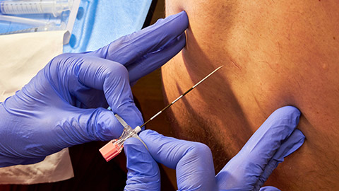
An epidural is a specialised route of administration for pain relief. A thin tube is inserted into the epidural space in the spine through which an appropriate analgesic, such as local anaesthetics, steroids and opioids, is administered. The analgesic blocks nerve signals in the spinal cord, resulting in temporary loss of sensation, usually in the lower part of the body, depending on where the epidural is inserted. This helps alleviate pain and discomfort, making it a commonly used method for managing pain during childbirth, certain surgeries, or chronic pain conditions.
The advantages of an epidural are that it blocks sensation to a specific area, allowing pain relief in a specific region without affecting the entire body. It is fast and effective pain relief, and the patient can be conscious and alert during a medical procedure and avoid potential complications from a general anaesthetic. The disadvantages of epidurals are that they can be associated with side effects such as low blood pressure, headache, and nerve damage.
Opioids: Oral
Morphine is an opioid analgesic. It binds to specific receptors in the central nervous system, known as opioid receptors. These receptors are primarily found in the brain and spinal cord.
Examples of oral morphine are:
- morphine sulphate SR (trade name m-Eslon®) capsules.
- morphine sulphate tablets (trade name Sevredol®).
Journal post
Pain: Oral Morphine
In this tūmahi (activity), you will research two morphine dose forms and share your findings in a journal post.
- Complete the following Documentation tool activity.
- Download and save the completed activity.
- Create a new journal post titled ‘Pain: Oral Morphine’.
- Upload the completed activity to the journal post and publish the post to ‘All course users.’
- Save the permalink to your Index of Journal Posts. Your tutor may review your answers and provide feedback, making it easier to check back!
Anxiety
Anxiety is a natural response within the nervous system that activates the body's "fight or flight" mechanisms in anticipation of a perceived threat. It involves the activation of specific regions of the brain which trigger the release of stress hormones like adrenaline. Anxiety can lead to physical symptoms such as increased heart rate, rapid breathing, and muscle tension.
Treatment for anxiety
The pharmaceutical treatment of anxiety may include medicines from the therapeutic class, benzodiazepines. Benzodiazepines work on the nervous system by enhancing the neurotransmitter gamma-aminobutyric acid (GABA) effects. GABA is an inhibitory neurotransmitter, meaning it dampens the activity of nerve cells in the brain. By increasing GABA's inhibitory actions, benzodiazepines help reduce overactivity in the brain, leading to a calming effect and relief from symptoms of anxiety.
Journal post
Anxiety: Lorazepam
Lorazepam is a benzodiazepine used in the management of anxiety. In this tūmahi rangahau (research activity), you will investigate this medicine and your findings in your journal. By reporting your research findings in your journal, you are creating useful resources for your future self!
- Create a journal post titled ‘Anxiety: Lorazepam’.
- Research to answer these questions:
- Explain the indications for use, therapeutic class and the action of lorazepam.
- For short-term use, what is the dose and frequency of lorazepam?
- What is the patient’s advice for lorazepam? Include cautions, side effects and how to manage them.
- Publish the post to ‘All course users’ as your tutor may review and provide feedback on your mahi (work).
- Save the permalink to your Index of Journal Posts.
Depression (Mate pāpouri)
Depression is a mental health condition with persistent feelings of sadness, hopelessness, and a lack of interest or pleasure in activities. Depression involves disruptions in the normal functioning of neurotransmitters such as serotonin, norepinephrine, and dopamine.
Treatment for depression
Treatment for depression may include the therapeutic class of antidepressants called Serotonin Reuptake Inhibitors (SSRIs). SSRIs block serotonin from reabsorbing, a process known as ‘reuptake’. This allows serotonin to remain in the synaptic cleft for a longer time, which is believed to contribute to the alleviation of depressive symptoms and the improvement of mood.
Journal post
Depression: Citalopram
Take a moment now to research a common SSRI, citalopram.
- Create a journal post titled ‘Depression: Citalopram’.
- Research to answer these questions:
- Explain the indications for use, therapeutic class and the action of citalopram.
- The typical dose and frequency of citalopram.
- What is the patient’s advice for citalopram? Include cautions, side effects and how to manage them.
- Publish the post to ‘All course users’ as your tutor may review and provide feedback on your mahi (work).
- Save the permalink to your Index of Journal Posts.
Migraines (Māhunga ānini)
You may be familiar with the nervous system disorder of migraines. You or someone you know may have experienced them. A migraine is a type of headache with intense, throbbing pain, often on one side of the head. It is typically accompanied by symptoms such as nausea, sensitivity to light and sound, and sometimes visual disturbances called auras. Migraines can significantly impact daily activities and may last for hours to days.
Treatment of migraines
There are many different treatments for migraines. Select each label or the (+) icon to read about the following treatments:
This therapeutic class of medicines may help treat migraines if taken as soon as an attack is felt. They interfere with the production of prostaglandins, which are chemicals that sensitise nerve endings and contribute to the perception of pain. By blocking these chemicals, NSAIDs can reduce pain signalling.
Rizatriptan belongs to the therapeutic class of medicines known as triptans, which act as a serotonin agonist. Serotonin agonists mimic the action of serotonin and directly affect the 5-HT receptors in the brain's blood vessels. This results in the constriction of intracranial arteries, relieving migraine symptoms.
Rizatriptan is generally a more effective treatment for an acute migraine attack than NSAIDs due to its more specific and targeted action to constrict blood vessels and due to triptan's relatively short-acting nature. When a migraine is relieved, a slow-release NSAID can be taken to prevent the recurrence of the headache.
Journal post
Migraine: Rizatriptan
Use your pharmacy technician knowledge and research skills to dispense rizatriptan in this scenario activity safely.
- Complete the following Documentation tool activity.
- Download and save the completed activity.
- Create a new journal post titled ‘Migraine: Rizatriptan’.
- Upload the completed activity to the journal post and publish the post to ‘All course users.’
- Save the permalink to your Index of Journal Posts. Your tutor may review your answers and provide feedback, making it easier to check back!
Epilepsy (Mate hukihuki)
Epilepsy is a neurological (nervous system) disorder characterised by recurrent seizures and a sudden burst of uncontrolled, disorganised electrical and chemical activity in the brain. A seizure prevents the brain from:
- interpreting and processing incoming sensory signals.
- controlling muscles.
You can read more about epilepsy on the Healthify webpage, including the different types of seizures: Epilepsy | Mate hukihuki.
Treatment for epilepsy
The most common treatment for epilepsy is the use of anticonvulsants, sometimes referred to as anti-seizure medication (ASM). How anticonvulsants work depends on the specific drug. Some, such as sodium valproate, are not fully understood but are thought to increase the activity of the neurotransmitter gamma-aminobutyric acid (GABA) in the brain. GABA is an inhibitory neurotransmitter that has a calming effect on the brain's electrical activity.
Journal post
Epilepsy: Sodium Valproate
Doing your own research and reporting on your findings helps you learn about the disorders and treatments of different body systems. As a pharmacy technician, this is vital learning that enables you to provide accurate and safe patient advice. The more you put into these research activities, the more you learn! On that note, it’s time to research and report back on sodium valproate…
- Create a journal post titled ‘Epilepsy: Sodium Valproate’.
- Research to answer these questions:
- What is the trade name of sodium valproate?
- What are the common side effects, and what can you advise a patient to do about them?
- Are there any medicines that should be avoided while taking sodium valproate?
- What cautionary and advisory advice should you provide when dispensing this medicine?
- What advice will you provide to both males and females about contraception while taking this medicine?
- Publish the post to ‘All course users’ as your tutor may review and provide feedback on your mahi (work).
- Save the permalink to your Index of Journal Posts.
Parkinson’s disease (Mate pākenetana)
Watch: 2-Minute Neuroscience: Parkinson’s Disease (2:01 minutes)
Parkinson’s disease (PD) is the second most common neurodegenerative disease behind Alzheimer’s disease. Watch this video to learn about what happens in the brain for people living with Parkinson’s, what effect this has on their ability to move, and what the most common pharmaceutical treatment is. Once you have watched this, answer the three quiz questions to ensure you’ve retained the key points.
Treatment for Parkinson’s disease
Medications used in the management of PD include the following. Select each label or the (+) icons to read more.
Action: These act in the brain to improve motor function by increasing dopamine concentration or enhancing dopamine's neurotransmission.
Example: Levodopa.
Action: These inhibit the action of the neurotransmitter acetylcholine. In PD, the effect of acetylcholine is stronger. Reducing the effect of acetylcholine can help to treat PD symptoms of rigidity, slowness of movement, tremors, speech and writing difficulties, gait, sweating, involuntary movements of the eyes and feeling depressed.
Example: Orphenadrine.
Attention Deficit Hyperactivity Disorder (Mate mauri rere)
ADHD is a neurodevelopmental disorder that affects the nervous system, particularly the brain. It is characterised by persistent inattention, hyperactivity, and impulsivity patterns that can impact an individual's daily functioning and development.
ADHD is believed to involve dysregulation of neurotransmitters, such as dopamine and norepinephrine, which play crucial roles in attention, focus, and impulse control.
Treatments for ADHD
In Aotearoa New Zealand, the pharmaceutical treatments for ADHD aim to improve concentration, reduce impulsiveness and promote calmness. These medications include:
- Methylphenidate
- Dexamphetamine
- Atomoxetine
Methylphenidate
Let’s take a closer look at methylphenidate.
Follow this link to read the New Zealand Formulary page on this medicine: Methylphenidate hydrochloride. Focus on the adverse effects, dosing regime, patient advice (read the information leaflet for patients) and CALs.
One of the side effects of methylphenidate is difficulty sleeping. This may be due to the effect of increased neurotransmitters, such as dopamine and norepinephrine, in the brain. These neurotransmitters affect wakefulness and alertness, and increased levels may interfere with the normal sleep-wake cycle.
As a pharmacy technician you may be asked for advice in managing this side effect. You already have some knowledge about ‘sleep hygiene’ from previous learning in this programme.
Sleep hygiene practices can be very helpful for patients and include:
- Having a regular sleep routine with regular bed and wake-up times.
- Having a balanced diet and exercise during the day.
- Creating an environment for sleep, such as a dark and quiet space and comfortable bedding.
Other advice in managing this side effect related to the medication include:
- Taking doses at the stated time.
- Not taking the dose too late in the day as its stimulating effects may persist into the evening, making it difficult for individuals to fall asleep.
- Talking to their doctor about sleep issues. Some people are more sensitive to the stimulant effects than others.
Self-directed learning activity
Mahi pai, excellent work this session. There was a lot of new information to digest; therefore, your self-directed activity this week is to review your learning.
You might like to solidify your knowledge of the nervous system disorders and treatments we have covered this week by reading, watching or listening to more information. You can add more information to your journal entries and/or any other notes you have been taking.
Useful resources for this include:
- Healthify website: Homepage
- Ministry of Health: Conditions and treatments section
- NZF: Homepage
- Trusted YouTube channels, e.g. Abraham The Pharmacist
We also encourage you to check that you have saved all the mahi you’ve done this week. For example, save your journal post permalinks to the Index of Journal Posts and save your completed Documentation tool activities to your device.
Kei runga noa atu! On to it! You’ve wrapped up another huge week of learning. Be proud of your efforts, and ensure you have a break to absorb your learning and recharge your energy before starting next week’s content.
