It is inevitable that you will come across information that includes references to the anatomy and physiology of clients and residents. These terms refer to the way that the body is structured or put together (anatomy), and the way that it works (physiology).
This chapter introduces you to the 11 body systems and how they work together to keep the body healthy. It will help you to understand and use basic medical terms used to describe body systems.
By the end of this chapter, you will understand:
- The basic structure and functions of cells, tissues and organs
- The body systems and how they work together
- Terminology that describes the normal structure, function and location of the major body systems.
Cells are the building blocks of our body. Every part of your body is made of cells.
Cells in your body do many different things. They can:
- Send messages between your brain and organs (nerve cells)
- Move body parts (muscle cells)
- Protect our body (skin cells)
- Carry oxygen and nutrients (blood cells)
- Store nutrients (bone cells)
Cells come in different shapes and sizes so that they can help an organ to do what it is supposed to do, but they have most of their basic structures in common. There are hundreds of different types of cells in the body.
The nucleus is like the brain of the cell. It controls what the cell does. It contains chromosomes which are like a blueprint. Chromosomes contain instructions that are passed on to new cells in cell division and cell replication.
Cells need food, water and oxygen to perform their functions. These pass through the cell wall. Most cells can repair themselves if they are damaged.

Tissues
Tissues are simply groups of cells of the same type.

- The muscle Tissue includes cardiac, skeletal, and smooth muscle
- The Epithelial Tissues are thin tissues covering all surfaces of our body that are exposed and form an external skin. For example, the inner lining of our mouth, our digestive tract, and hollow parts of our organs such as our heart, eyes, ears, and lungs.
- The nervous tissue is the main tissue of our nervous system which regulates and monitors the functions of our body.
- Connective tissue serves to connect, bind and support all the other tissues in our body.
Activity
Watch this YouTube video from Fun Science Demos and then answer the following questions:
Q1. Where in your body can you find cells?
Q2. What is the difference between cells, tissues, and organs?
An organ is made up of tissues to perform a particular function. Our body has 79 organs. They are connected to other organs through networks of blood vessels and nerves. The heart, kidneys and liver are all examples of organs.
This section will look at some of the major organs and the systems they belong to.
Describing Organs
Organs can be described in relation to their structure and their function.
Structure
This refers to how the organ or system looks like, what it is made of and what form it takes. For example, the heart is made of muscle cells and consists of four chambers connected to our major blood vessels.
Function
This refers to what the organ or system does, what its purpose is and what job it performs. For example, the heart’s function is to pump blood into the main blood vessels so that it can travel around the body and deliver oxygen to the cells.
The Body Systems
We group organs together depending on their overall functions. A group of organs that work together for a common function is called a system. We have 11 systems in the body.
- Digestive system
- Nervous system,
- Special senses
- Integumentary system
- Reproductive system
- Urinary system
- Lymphatic system
- Cardiovascular system
- Endocrine system
- Immune system
Watch
Watch this YouTube video by National Geographic called Human Body 101 for an overview of the body systems:
The Body Cavities
Many of our systems and organs sit inside cavities. Here is a list of the cavities in the body:
| Cranial cavity | Inside the head, holding the brain. |
|---|---|
| Vertebral canal | The space inside the vertebrae. Contains the spinal cord. |
| Thoracic cavity | The large cavity in the chest that holds the heart and lungs. |
| Pleural cavity | The spaces that surround both lungs, filled with fluid. |
| Pericardial cavity | The fluid-filled space that surrounds the heart. |
| Abdominal cavity | The cavity in the abdomen that contains the liver, stomach, spleen, small intestine and most of the large intestine. |
| Pelvic cavity | The cavity in the pelvic area that contains the bladder, some of the large intestine and the internal reproductive organs. |

- Brain
- Oesophagus
- Lymph nodes
- Liver
- Kidneys
- Large intestine
- Muscle
- Lungs
- Blood vessels
- Stomach
- Small intestine
- Bone
- Joint
The Nervous System
The nervous system is made up of the brain, the spinal cord and nerves.
The nervous system is made up of the brain, the spinal cord and nerves.
- The brain is located in the skull, inside the cranial cavity.
- The spinal cord runs through the vertebral column of the spine.
- The nerves travel to every part of our body.
The brain is the body’s control centre. We use our brain to create both conscious and unconscious actions in the rest of the body.
The brain sends messages through the nerves to all parts of the body via the spinal cord.
The nerves are like an electrical cord. The messages travel along chains of nerve cells where they act on the body part in the way the brain directs.
Example
Conscious actions include muscle movement. For example, when we want to pick up a pen, our brain sends a message to our arm and fingers telling the muscle how, when and where to move.
Unconscious actions are the body functions that we do not consciously control. For example, the brain sends a message to the diaphragm which moves up and down, allowing us to breathe
Watch
Watch this YouTube video called A Journey Through Your Nervous System by BRIGHT SIDE:
The Cardiovascular System
Consists of the heart and a network of blood vessels that spread to all parts of the body
Pumps blood around the body to provide oxygen, hormones and nutrients to the cells
The heart is in our thoracic cavity, while the blood vessels and capillaries travel around the entire body
The cardiovascular system is sometimes called the circulatory system.
The cardiovascular system consists of:
Heart
A muscular organ which pumps blood around the body

Arteries
Carry oxygenated blood away from the heart to the rest of the body.

Veins
Carry blood back to the lungs to refuel with oxygen before returning it to the heart.
Capillaries
Tiny vessels that lead from the veins to the tissues and cells.

Blood
Contains blood cells which carry oxygen and other components around the body

Watch
Watch this YouTube video by National Geographic called Heart 101 about how the heart does its work:
The Respiratory System
- Includes air passages, the lungs and breathing muscles.
- Our lungs and bronchi look a bit like an upside down tree.
- Allows us to breathe in air, providing oxygen to the body tissues.
- Removes waste such as carbon dioxide.
The organs of the respiratory system are contained in our thoracic cavity

- Nasal cavity
- Nostril
- Epiglottis
- Pharynx
- Larynx
- Trachea
- Primary bronchus
- Right lung
- Pleural cavity
- Left lung
- Diaphragm
All of our body cells and tissues need oxygen. The respiratory system helps us to take oxygen from the air so that it can be used by our cells.
Example
Some systems have higher demand for oxygen than others. Our brain needs a great deal of oxygen to perform its functions.
Watch
Watch this YouTube video titled Lungs 101 by National Geographic about how the lungs exchange oxygen with the blood cells:
Here is the simple process that allows us to breathe in and out:
Inhalation
When the diaphragm muscle move down, air enters our respiratory system via the mouth and nose. This is called inhalation. Our respiratory system is lined with tiny hairs that trap dust and other particles to help purify the air.
It passes through the pharynx and into the bronchi. These branch into bronchioles, where the air enters the lungs. The lungs consist of tiny air sacs called alveoli. These are lined with capillaries. The oxygen passes into these capillaries and enters the blood stream. The air is now deoxygenated; this means that the oxygen has been removed.

Exhalation
Carbon dioxide enters the lungs from the blood stream and is passed out of the lungs via the trachea when the diaphragm muscle moves up. This is called exhalation.

The Musculo-Skeletal System
- Made up of bones and muscles connected by tendons and ligaments
- These are connected by tendons, ligaments, cartlilage and joints
- Helps us to stay upright
- Helps us to move
- Helps organs to do their work
Every area of our body contains muscles and bones

The musculo-skeletal system is made up of the muscles and bones (skeleton).
The Skeleton
The skeleton is made up of 206 bones. These give our body its shape and help us to stand and sit upright. Some bones, such as the ribs, protect our organs. Other bones, such as the bones in our arms, legs and neck help us to move.
Bones have other important functions that you may not know about. Our larger bones, such as our leg bones, make blood cells inside the bone marrow. The bones also store important minerals such as calcium.
Muscles
The muscles allow movement. They also help us to stay upright and produce heat. There are three types of muscles:
- Cardiac Muscle
- This muscle type is only found in the heart. It has its own electrical impulses, just like a battery and it keeps moving even if the connection is lost with the brain

- Skeletal Muscle Tissue
- This is attached to our bones. We have conscious control over skeletal muscle, for instance, we can move.

- Smooth Muscle Tissue
- This muscle is contained in many organs such as our intestines that need smooth muscle to perform their function. We have no control over our smooth muscles.

Watch
Watch this YouTube video by Dr Matt & Dr Mike called Muscle Tissue about how the muscular system works:
The Endocrine System
Made up of glands which secrete hormones.
- Send chemical messages around the body.
- These messages have many different functions.
Glands are located in many different parts of the body, including the brain.

- Hypothalamus
- Pituitary gland
- Pineal gland
- Thyroid and parathyroid glands
- Pancreas
- Adrenal glands
- Placenta
- Thymus
- Ovary (in female)
- Testicle (in male)
Hormones send chemical messages to the body’s organs to tell them to do something. We have hormones that send messages to control reproduction, growth, digestion and many other functions.
Example
The pancreas secretes a hormone called insulin. This hormone tells our body to decrease the levels of glucose (sugar) in our blood when they become too high. Diabetes happens when there is a problem with the manufacturing of this hormone.
Glands are the organs that produce and release hormones.
Here are the main glands of the body and the actions they control:
| Name of gland | Where is it? | What does it do? |
|---|---|---|
| Hypothalamus | At the base of the brain | Controls our sleep, appetite, weight, thirst, blood pressure, sex drive, memory and mood |
| Pituitary gland | In the base of the brain | Controls blood pressure, blood sugar levels, stress responses, menstruation, sperm production, bone and muscle growth |
| Pineal gland | Near the centre of the brain | Produces melatonin which controls our sleep cycle |
| Thyroid gland | In the front of the neck | Controls our metabolism, energy levels, body temperature, heart rate and blood flow |
| Parathyroid glands | In the neck behind the thyroid gland | Controls calcium levels in the blood |
| Adrenal glands | Above the kidneys on each side of the body | Produces adrenaline, cortisol and testosterone Controls stress responses, blood pressure, energy, sex organs and heart rate |
| Pancreas | Behind the stomach, under the liver | Releases insulin to control blood sugar levels |
| Ovaries | On each side of the uterus in the pelvis | Releases oestrogen, progesterone and testosterone Controls female characteristics, releasing eggs |
| Testes | In the scrotum, behind the penis | Produces testosterone, controls male characteristics and sperm production |
Watch
Watch the following YouTube video called Overview of the Endocrine System by Dr Matt & Dr Mike:
The Digestive System
A long tract that includes the mouth, oesophagus, stomach, liver and intestines
- Breaks down food into smaller, liquid nutrients
- Allows nutrients and water to be used by the cells in the body
Leads from the mouth to the anus. The intestines are located in the abdominal cavity
We take food into our mouth. Our tongue and teeth help us to move the food and break it down into smaller pieces so that it is ready for swallowing.
We swallow the food and it passes into our pharynx at the back of the throat. A small flap of muscle covers over the trachea (windpipe) to prevent food from passing down into our lungs. Instead, it passes to another tube called the oesophagus. This is a muscular tube that pushes the food down towards the stomach. In the stomach, the food is churned and mixed to become a liquid. The stomach releases acids, which help to break the food down further.
The gallbladder releases bile into the stomach. Bile acts like detergent; it helps to break down fats and oils.
Once the food is liquified, it then moves into the small intestine (small bowel). This is where the nutrients in the food leave the digestive system. They pass through the walls into the capillaries and blood stream where they travel to the cells. The small intestine is very long.
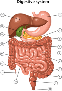
| Number | Organ |
|---|---|
| 1 | Stomach |
| 2 | Pancreas |
| 3 | Left splenic flexure |
| 4 | Transverse colon |
| 5 | Jejunum |
| 6 | Descending colon |
| 7 | Sigmoid flexure |
| 8 | Sigmoid colon |
| 9 | Rectum |
| 10 | Anus |
| 11 | Ileum |
| 12 | Appendix |
| 13 | Cecum |
| 14 | Ascending colon |
| 15 | Right hepatic flexure |
| 16 | Duodenum |
| 17 | Gallbladder |
| 18 | Liver |
This is important because the nutrients move out slowly as the food passes through. If the small intestine was shorter, we would not be able to take all of the important nutrients that we need from our food.
The large intestine is a larger, shorter tube that helps us to remove water from the remains of the food. Water travels through the wall of the large intestine (large bowel) and into the blood stream. At the beginning of the large bowel the food (now faeces) is watery and liquid. By the time it enters the lower part of the bowel, it is solid faeces. The faeces now contains all of the waste that the body does not want or need. These must be removed from the body.
The solid faeces sits in the rectum where it is stored until we are ready to go to the toilet. Nerves in the rectum are stimulated by the presence of faeces, and we feel the urge to push. Muscles help us to push the faeces out of the body.
Watch
Watch this YouTube video by TEDEd called How your digestive system works - Emma Byrce:
The Urinary System
Consists of the kidneys, ureters, urethra and bladder
Filters the blood to remove excess water and waste products, which are then stored in the bladder and excreted as urine
The kidneys are located towards the back (posterior) and the bladder and urethra is contained in the abdominal cavity

- Renal Artery
- Kidney
- Bladder
- Renal vein
- Kidney content
- Urethra
Blood passes through the kidneys. These are two bean-shaped organs that filter the blood, drawing out water, minerals and other substances that the body does not need. This takes the form of urine.
The urine passes down through two long tubes called ureters to the bladder. The bladder is a little like a storage container; urine is stored there until we are ready to go to the toilet or until it is too full to hold more.
When the bladder is full, a message is sent to our brain to tell us that we need to pass urine. When we are ready, the brain sends a message to a sphincter, a muscle that sits at the bottom of the urethra. The sphincter relaxes and the urine passes through the urethra and out of the body.
Note
The male and female urinary systems are very similar. One of the only differences is the length of the urethra. In males, the urethra is longer because it passes through the penis. Females are more prone to urinary tract infections because germs do not have to travel far through the shorter urethra to make it to the bladder.
Watch
Watch this YouTube video by Dr Matt & Dr Mike called Renal Function about the urinary (renal) system:
The Reproductive Systems
- Males and females have different reproductive systems.
- The female reproductive system is made up of the uterus, ovaries, fallopian tubes and vagina.
- The male reproductive system is made up of the penis, testes, epididymus and vas deferens.
Both systems contribute to reproduction and birth.
The reproductive organs are located in the pelvic cavity.
The Female Reproductive System
The female reproductive system contains the organs required to conceive and have children. Females are born with many egg cells at puberty. The ovaries develop and produce eggs, ready to be fertilised by the male sperm. Menopause, the end of this process, usually occurs between the ages of 45 and 55. Many older women experience changes after menopause because of the reduction in hormones in their body. This can lead to physical changes such as loss of bone density and mood changes.
Watch
Watch this YouTube video by Dr Matt & Dr Mike titled Female Reproductive System:

- Uterus
- Fallopian tube
- Vagina
- Cervix
- Ovary
The Male Reproductive System
The male reproductive system is focused around the production of sperm and delivering sperm to the female during sex. Sperm is produced in the testes.
The prostate gland produces fluids that feed and protect sperm cells. As men age, this gland can commonly enlarge and cause problems, including urinary problems and cancer.
Watch
Watch this YouTube by Dr Matt & Dr Mike called Male Reproductive System:
The Integumentary System
Consists of skin, nails and hair
Provides a protective covering for the body
Helps to regulate body temperature

- Hair
- Sweat pore
- Sweat gland
- Vein
- Artery
- Adipose tissue
- Subcutaneous layer
- Hypodermis
- Derma
- Epidermis
The skin is the body’s largest organ. It contains blood vessels, sweat glands and very dense nerve endings. Skin consists of two distinct layers:
The epidermis
- The epidermis is the outer layer of skin
- It contains epithelial calls. These supply a waterproof layer to the body
- The cells continuously divide, die and shed
The dermis
- The dermis is the layer under the skin
- It is made of connective tissue and collagen fibers
- The dermis contains blood vessels and nerve endings so that our skin can feel touch, pressure, heat, cold and pain
- The dermis also contains hair follicles, sweat glands and sebaceous glands
Watch
Watch this YouTube video by National Geographic called What is Skin?:
Sebaceous glands produce sebum, an oil that helps to waterproof and moisturise the skin and hair. Sebum also controls the growth of disease-causing microorganisms on the skin.
Sweat glands produce sweat, which contains water and salts.
Hair helps to insulate and protect the body. As we age, our hair tends to thin, increasing our susceptibility to cold. Clients who have little hair on their head, such as balding men, might need extra head coverings in cold weather.
Eyelashes and eyebrows protect eyes from water, dirt and infections.
Note
Melanocytes are cells that produce melanin and protect us from UV rays. People with whiter skin are at more risk of sunburn than people with darker complexions, because they have less melanin.
People with darker skin need more time in the sun to absorb adequate Vitamin D. They are more at risk of Vitamin D deficiency than people with lighter skin.
The Lymphatic System
- Consists of lymph nodes, the lymph vessels and lymph fluid
- The spleen and thymus are also part of the lymphatic system
- Filters bacteria from the tissues
- Drains excess fluid from the tissues and returns it to the veins
Lymph nodes are located at major body junctions, such as armpits and the groin. Lymph vessels run alongside the blood vessels all over the body
The lymphatic system is a network of tiny vessels that travel through the body to help drain fluid from our tissues and contributes to fighting infections.
Lymph is a clear fluid that contains white blood cells. The white blood cells fight infections in all of our body’s tissues and organs.
Lymph vessels are vessels that carry the lymph fluid to the organs and tissues. They also drain away extra fluid that the capillaries cannot reach. This fluid is drained from the lymph vessels into the surrounding capillaries and veins.
Lymph nodes are also called lymph glands. The lymph vessels travel through them at key points where they trap disease causing bacteria and microbes.
The spleen filters old and damaged blood cells and removes and destroys infections.
The thymus filters the blood to keep it clean and also produces lymphocytes.

- Palatine tonsil
- Axillary lymph nodes
- Spleen
- Thoracic duct
- Inguinal lymph nodes
- Cisterna chyle
- Right lymphatic duct
- Thymus
- Cervical lymph nodes
Watch
Watch this YouTube video titled The Lymphatic System Overview, Animation by Alila Medical Media:
Example
When lymph nodes and lymph vessels are damaged, the body parts nearby can become swollen with fluid that is not able to be drained away. For example, during breast cancer surgery, the surgeon may need to remove lymph nodes and vessels in the thoracic area and neck. This can mean that the person is more prone to swelling in the arm on that side of their body, because the lymph fluid is not able to be drained back to the blood vessels effectively.
The Immune System
The immune system is made up of:
- The lymph vessels
- The spleen and the thymus
- White blood cells, bone marrow and antibodies
Fights infections such as viruses and bacteria
- White blood cells and antibodies travel in the blood
- The lymphatic system extends over the whole body alongside the veins and arteries
- The spleen is in the left upper abdomen. The thymus is located behind the breastbone (sternum)

- Tonsils and adenoids
- Thymus
- Lymph nodes
- Spleen
- Appendix
- Peyer’s patch
- Bone marrow
White blood cells are made in the bone marrow and work with antibodies to attack foreign bodies. White blood cells include lymphocytes known as a memory cells. Memory cells recognise and destroy certain disease-causing organisms more quickly when it recognises them before they can multiply. This is why it is uncommon to develop some infections, such as chicken pox or measles, more than once.
When a person has a blood test that shows high white blood cell levels, this can mean that there is an infection present that the body is trying to fight. High leukocyte levels in a urinalysis test, (which you will learn more about in chapter 2) can also be a test to show that there is infection present that the body is trying to fight.
The flu and the common cold, however, have many different strains of the same type of virus, so the lymphocytes do not recognise the new strains.
Bone marrow is found inside your large bones. It produces red blood cells, white blood cells and platelets.
The immune system works with many other parts of the body too, to help protect us from infection.
Here are some examples:
- The lymphatic system
- The skin, saliva and tears secrete enzymes that kill bacteria
- Mucous and tiny hairs in the respiratory system trap foreign particles such as infections, allowing us to remove it via coughing
- The digestive system contains antibodies and the acid in the stomach kills most bacteria
- When a person develops an infection, a fever develops. This means that the person’s body temperature rises, killing infections that prefer normal body temperatures
Note
Vaccines trick the body into producing antibodies to fight a specific virus. If they are later exposed to the actual virus, they will not get sick because their body will recognise it and attack it.
People who work in residential aged care must be vaccinated against the flu and COVID-19, and there are similar rules for people working in other community service workplaces.
Sensory Systems
Our sensory systems are made up of the special senses.
Read more about it here
Read the above article and describe each of the following senses:
- Taste
- Smell
- Equilibrium
- Hearing
- Sight
The organs responsible for helping us to use our senses are highly specialised. This section outlines some of these structures.
The Eye

- Sclera
- Cornea
- Iris
- Pupil
- Macula
- Ciliary body and muscle
- Conjuctiva
- Retina
- Vitreous body
- Reginal blood vessels
- Optic nerve
- Macula
The eye lets images pass through and focuses it on the back of the eye, called the retina. The retina is covered with cells called rods and cones. These cells are light sensitive; they pick up colour, shapes and light, and send the image to the brain via the optic nerve. The right and left eye see a slightly different view. The brain combines the two views and produces a three-dimensional image.
Here are some of the important parts of the eye:
The iris is a muscle that contracts and dilates the pupil to control the amount of light entering the eye. In dark light, the pupil widens to let in more light. In bright light, it constricts.
The conjunctiva is the thin clear membrane that covers the eye.
The sclera is the medical term for the whites of the eye.
The cornea is a clear lens that helps us to focus.
The lens sits behind the iris and provides short focus.
Tear ducts lubricate the eye to stop it from drying out and flushes away dust and bacteria.
Note
- As we age, the muscles that control focus and vision begin to deteriorate.
- This can lead to short-sightedness or longsightedness.
- Cataracts happen when the lens becomes cloudy. Other common eye conditions include glaucoma and macular degeneration. These conditions become more common as we age.
- Your eyes take three times as long to adjust to the dark in your seventies as they do in your twenties.
Watch
Watch this YouTube video by National Geographic called Eyes: The Windows to Your Health:
The Ear

- Semicircular canals
- Cochlea
- Vestibular nerve
- Cochlear nerve
- Eustachian tube
- Tympanic cavity
- Tympanic membrane (Eardrum)
- Cartilage
- Auricular lobule (Earlobe)
- Concha
- Antihelix
- Triangular fossa
- Scapha
- Helix
- Temporal muscle
- Temporal bone
- External acoustic meatus (Ear canal)
- Malieus
- Incus
- Stapes
The ear provides hearing and balance. It is made up of three different parts:
The outer ear
- This part of the ear collects sound waves
- It is coated with wax, which lubricates, fights infection and clears dead skin
The middle ear
- This part of the ear contains the eardrum and three small bones
- The eardrum vibrates and causes the tiny bones to move
- Sound is carried through these bones to the cochlea in the inner ear
The inner ear
- Here the sound waves are passed over thousands of tiny hairs and converted to electrical impulses and sent to the brain
- The brain interprets the sound
- The vestibular system of the inner ear also controls balance
When you are working with older people and people with disabilities, you will often read and hear information that relates to parts of the body. Sometimes you will need to share this information with other workers and the client or resident.
Disabilities and medical conditions often involve parts of the body that are described by the affected organs or systems.
Example
A person with a spinal cord injury might talk about symptoms involving the nerves of their body. When you understand the basic parts of the nervous system, you will know more about the person’s needs for support. You will also understand the importance of reporting problems with the functioning of this system.
You might see or hear information about the body:
- When the client or resident talks to you about their condition, or about symptoms they are currently experiencing, such as abdominal pain and constipation
- In charts used to collect information, such as bowel charts, temperature charts and fluid balance charts. The next section of this resource will describe the use of some of these charts and your role in using them
- When you are given a handover about the person, such as when you are told that the person has a brain injury
- When a medical or allied health professional writes notes or instructions to you and other staff, such as the position of a muscle that needs to be exercised
- When you are sharing information about healthy lifestyle changes, such as a healthier diet or support to quit smoking
- When you are reading file notes written by other staff, such as about the condition of the person’s skin
The Interrelationships of Organs
The language of anatomy and physiology is very specific. In many cases, it can be easy to learn the main parts of the medical language. Here are some examples of terms used to describe where organs and systems are placed in relation to each other.
| TERM | MEANING | EXAMPLE |
|---|---|---|
| ANTERIOR | At the front of | The anterior cruciate ligament (ACL) is a ligament at the front of your knee joint |
| POSTERIOR | At the back of | The posterior tibial tendon is a tendon behind the lower part of tibia, the bone that joins the ankle to the lower leg |
| LATERAL | To the side of the body | The heart is in the left lateral side of the chest |
| MEDIAL | Near the middle of the body | The spinal cord is in a medial position in the body |
| SUPERIOR | Above another organ | The lungs are superior to the diaphragm |
| INFERIOR | Below another organ | The intestines are inferior to the stomach |
| SUPERFICIAL | Close to the surface of the body (near the skin) |
The sweat glands are superficial organs |
| DEEP | Away from the surface of the body | The bones are deep structures |
| PROXIMAL | Close to another organ or structure | The heart is proximal to the lungs |
| DISTAL | Further away from another organ | The metatarsals (foot bones) are distal to the ankle |
How Organs and Systems Work Together
Our organs and body systems do not work in isolation from each other. Every organ and system is dependent on other organs to complete its work.
For example:
- The lungs depend on the heart and blood vessels to transfer oxygen into the bloodstream
- The urinary system relies on the nervous system to send messages to the bladder
- The digestive system relies on the smooth muscles of the intestine to push and expel faeces towards the rectum
- The bones need minerals that can only be supplied by the digestive system.
When a person develops an illness or dysfunction in one part of the body, this can have an effect on the functioning of other organs.
Example
Diabetes is a disorder of the pancreas. It happens when the pancreas is unable to produce adequate amounts of insulin. Diabetes causes the person’s blood sugar levels to rise too high for healthy functioning because insulin is not available to help control the sugar levels.
Because there is too much sugar in the bloodstream, most other systems and organs are affected.
Cardiovascular system:
- Glucose can block the capillaries of the toes. This can reduce the blood flow, leading to problems with circulation. It can also cause gangrene which is the death of the tissues
Sensory system:
- Glucose builds up in the tiny capillaries of the eyes. This can lead to these capillaries becoming blocked and damage to the eyesight can result
Nervous system:
- Glucose can block the small vessels in the brain. This can cause damage to the nervous tissue leading to stroke
- Glucose can block the nerves. This can mean that the person can lose feeling or sensation in the nerves of their toes, feet, and other parts of the body. This is called peripheral neuropathy
- When the person cannot feel sensation in their feet, they may have trouble walking safely because they are more likely to trip. This can lead to a higher risk of falling and broken bones
Activity
Read this article before considering the two (2) questions that follow.
- What is the function of the central nervous system?
- What is the function of the & peripheral nervous system?
Use the following questions to check your knowledge.
- Give three examples of body cavities.
- Describe the structure of the urinary system.
- Explain the functions of the respiratory system.
- Describe the position of the rectum using medical terms.
- Explain the difference between an organ and an organ system.