Joints or 'articulations' are formed at the point where two bones meet. They are the connection between bones in the body and link the skeletal system as a functional whole system. Without joints, our skeleton would be a rigid deck structure unable to move. Have you ever been unfortunate enough to have a limb in a cast? If so, you know how this limited functional movement.
Joints and connective tissue hold the bones of our skeletal system together while still allowing some degree of movement.
A joint is a point of contact:
- between two bones.
- between bone and cartilage.
- between bone and teeth.

Joints have 2 main functions:
- Hold the skeleton together
- Give the skeleton mobility
In this topic, we focus on the names, structure, function and classification of joints within the skeletal system. You will learn:
- The science behind names
- Joint types and classifications
- Synovial joints
- Cartilaginous joints
- Fibrous joints
- Common joint conditions
- Exercise and joint considerations
In anatomy and physiology, you will read terms in both Latin and Greek as well as English. This can feel overwhelming. But, if we break down words into smaller parts, they become more familiar.
Typically, the scientific joint names are derived from the names of the bones involved. So looking at the names help us gather information about the structure, function, and location of the joint.
With practice, you will soon be able to easily 'translate' these names. This will support your understanding of anatomy and physiology. When this knowledge is combined with knowledge of muscle tissue, structure, and function, you will have a solid understanding of:
- where muscles and bones are located in the human body.
- which muscles are in a specific area.
- what bones are involved.
- which joints are present.
- how these all work together to provide particular movements.
Translating joint names
The scientific joint names are derived from the names of the bones involved. Let's break down "sternoclavicular joint".
- "sterno" = the sternum
- "clavicular" = the clavicle
So the sternoclavicular joint is where the clavicle meets the sternum. In the image, you can see this also applies to the acromioclavicular joint. It is located between the acromion and the clavicle.

- Sternoclavicular joint
- Clavicle
- Acromiaclavicular joint
- Acromion
- Concaid process
- Sternum
- Scapula
- Humerus
- Supraspinatus m.
- Subscapularis m.
- Infraspinatus m.
- Teres minor m.
Can you figure out which bones are involved in the following joints? Expand the accordion to check.
The tibiofemoral joint is where the femur meets the tibia. In this situation, the joint (synovial) is made complete by the patella (knee cap). Note, the patella completes the knee structure and is not classified as a component of a synovial joint.
The glenohumeral joint is where the humerus meets the glenoid cavity of the shoulder.
Of course, there are more common names for the key joints of the body.
- Hip joint: Iliofemoral joint (involving the ileum and the femur)
- Knuckle join: Metacarpophalangeal (involving the metacarpal bones and the phalangeal bones)
- Elbow joint: Humeroulnar joint (involving the humerus and the ulna)
- Ankle joint: Talocrural joint (involving the tibia, fibula and the talus (heel bone))
Sometimes it pays to be specific. As you've now seen, there are multiple joints that we could call a 'shoulder joint'. The language you use with clients, colleagues, and other health professionals will differ.
The skeletal system contains different classifications of joints based on structural composition and function (degree of movement).
Structural classification
The structural classification of joints is based on the dominant type of connective tissue.
- Synovial joints
- Cartilaginous joints
- Fibrous joints
Functional classification
The functional classification is based on the amount of movement permitted.
- Diarthrosis (freely moveable)
- Amphiarthrosis (slightly moveable)
- Synarthrosis (immovable)
How do structural and functional classifications relate?
The two classification schemes correlate.

Synovial joints are diarthrosis
They move freely. These can be further broken down into 6 groups based on how they move:
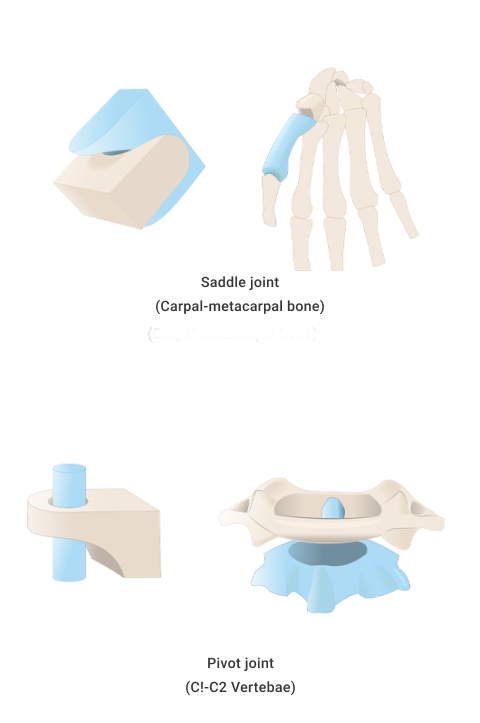
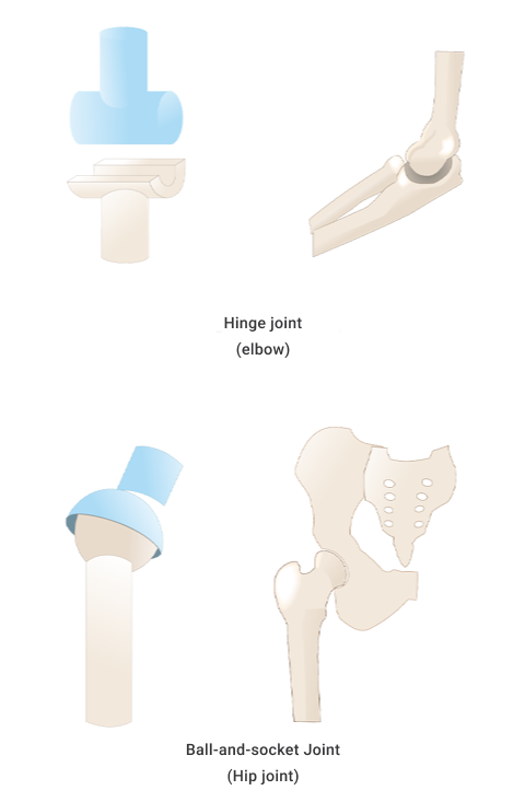
- Ball and socket joint
- Hinge joint
- Pivot joint
- Gliding (plane) joint
- Saddle joint
- Condyloid (ellipsoidal) joint
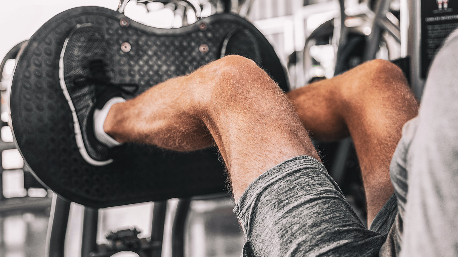
As a personal trainer, synovial joints are the joints you should become most familiar with. The risk of injury is generally greater at these joints due to the greater range of movement and subsequent reduction of the strength of the joint. These are the joints where movement happens, so knowledge of these joints and how they move is essential to exercise prescription. Synovial joints are where we will focus the most when studying joints for personal training.
Cartilaginous joints are amphiarthrosis
They are slightly moveable. Movement is needed but only to a certain degree. An example of this type of joint would be intervertebral discs in the vertebral column, where individual vertebrae are separated by cartilage, allowing some slight movement.
Fibrous joints are synarthrosis
They do not move. These joints are held together by tough tissue (specifically fibrous connective tissue), which develops during childhood. The cranium is an example of a fibrous joint (Physiopedia, n.d.).
Compare the images in the following slider. Look at:
- The knee joint in the first image
- The vertebral column or spine in the second image
- The cranium in the third image
Do the movements yourself. Are the joints moving freely, moving slightly, or immovable?
The primary purpose of synovial joints is to prevent friction where two or more bones meet. All synovial joints are diarthrosis joints, but the extent of movement varies and is often limited by the connecting ligaments. Nearly all joints of the limbs and most joints of the body are synovial joints (Physiopedia, n.d.).
Structure
A synovial joint has a synovial cavity between the bones. This cavity is filled with synovial fluid. The synovial fluid:
- allows smooth, frictionless movement.
- provides nutrients.
- removes wastes.
The synovial fluid acts as a lubricant, like oil in an engine, allowing all parts of the joint to move against each other smoothly. A synovial cavity allows significant movement of the joint.
Synovial joints also contain hyaline cartilage, known as articular cartilage. Articular cartilage provides shock absorption and reduces friction.

Typical structural components of synovial joints
Synovial fluid is produced by the synovial membrane and housed within the cavity. Synovial fluid is a semi-thick, clear/pale yellow fluid (which resembles the consistency and appearance of an uncooked egg white). This fluid forms a thin covering over the surface of the articular capsule.
The articular cartilage lines the ends of the bones but does not join them.
A fibrous joint capsule surrounds the joint to join the two articulating bones and enclose the synovial cavity.
The synovial membrane (synovium) secretes synovial fluid.
Accessory ligaments are present in the region of the synovial joint, e.g., the medial and lateral ligaments of the knee. They are not part of the synovial joint directly, but they support the joint.
Types of synovial joints

| Type of joint | How it moves | Example(s) |
|---|---|---|
| Ball and socket | Ball and sockets are due to the end of one bone having a 'ball' like end and the other bone having a 'socket' that the ball sits in. They move in multiple planes; for example, to flex, extend, rotate, abduct, adduct, and in circumduction. | Shoulder and hip |
| Hinge | A hinge moves in one plane only, like a door hinge. | Elbow and knee |
| Pivot | A pivot is a pure 'rotating' joint. | Neck, between C1 and C2 |
| Saddle | A saddle is when the end of one bone is like a concave 'saddle' that the other bone sits over, like two legs on a riding saddle. It allows a substantial range of movements. | Fingers and thumbs |
| Gliding (plane) | Gliding occurs when short bones glide along each other in all directions (but not far). Think of it like sliding two coins together between your fingers. | Intercarpal |
| Condyloid (ellipsoidal) | Condyloid refers to shallow ball and socket joints that can do everything a ball and socket joint can do except rotate. | Wrist |
Name that joint!
Practice labelling the joints to get familiar with the names. Use the exact spelling as the list provided in the activity. For example, "Ball-and-socket" contain hyphens.
Movements
The movements provided by synovial joints include:
- Flexion and extension
- Abduction and adduction
- Rotation
- Supination and pronation
- Lateral flexion
- Elevation and depression
- Protraction and retraction
- Ankle dorsiflexion and plantar flexion
Review Movement Terms in Anatomical Terminology if you need a refresher.
Move your synovial joints corresponding to those circled in the image. Identify the type of joint and the movement(s) for each joint.
Based on your knowledge of movement terms and how the different synovial joints move, can you match the movements to the joint type?
Cartilaginous joints contain no synovial cavity. The articulating bones are joined by cartilage (hyaline or fibrocartilage). They provide more movement than fibrous joints but less than synovial joints. The joints between the ribs and the sternum (breast bone) are examples of cartilaginous joints. They are represented in blue in the following image.

Structure
Cartilaginous joints occur where the bones are bound by cartilage (either hyaline or fibrocartilage) which permits little to no movement. Cartilaginous joints lack a synovial cavity.
Two types of cartilage can join bones via a cartilaginous joint:
Softer and with more give. Glassy appearance, often with a bluish hue. Think of the gristle on the end of a chicken drumstick bone.
Tougher, denser, more rigid cartilage.
Structurally, there are two types of cartilaginous joints:
- Synchondroses (primary cartilaginous joints)
- Sympheses (secondary cartilaginous joints)
Symphyses
Symphyses (the plural of symphysis) may have either hyaline or fibrocartilage. These joints are slightly mobile. E.g., the pubic symphysis within the pelvis provides strength and stability, plus flexibility and movement during times such as pregnancy and childbirth.

Synchondroses
Synchondroses refers to where the articulating bones are connected via hyaline cartilage. For example, the epiphyseal plate (also known as the growth plate) within long bones.

Fibrous joints lack synovial cavities. Bones are held together by dense, irregular connective tissue composed of collagen fibres that emerge from the matrix of one bone into the matrix of the next. The bones do not have a joint cavity between them and instead are directly connected to each other. The amount of movement depends on the length of the connective tissue. Some may be slightly moveable, but in general, most fibrous joints are immovable.
Fibrous joints fall into 3 categories.
- Sutures
- Syndemoses
- Gomphoses
Examples of fibrous joints include the joints which hold the teeth in their sockets, and joints holding the bones of the skull together.

Sutures
Sutures are typically found between the bones of the skull. In older adults, these sutures are immovable. However, in children and infants, they are slightly moveable. A baby's skull has 'soft spots' when born. It is this softer skull that allows movement and aids in childbirth. As the baby develops, we see the connective tissue turn to bone, and the skull grows and solidifies.

Synostosis
Synostosis is a type of suture that, as a person develops into adulthood, changes its structural composition, and some sutures are replaced by bone. When this occurs, this joint is no longer referred to as a suture but a suture that is now synostosis, where two bones have completely fused into one. Examples include:
- Bones of the skull
- Shafts and heads of long bones
- First rib to the sternum
Syndesmoses
Syndesmoses are immovable to slightly moveable. Within a suture, there is no space between the articulating bones. However, within a syndesmosis, there is more distance and volume of dense irregular connective tissue between the articulating bones of a syndesmosis.
The bones are held together by an interosseous membrane. There are two main interosseous membranes within the human body, between the tibia and fibula (shin bones) of the leg and the radius and ulna of the forearm.

Gomphoses
These are immobile joints between the teeth and their sockets.

As we age, we will likely experience some wear and tear, inside and out. Within the human skeletal system, degenerative conditions do present themselves. Aging usually impacts functions such as:
- synovial fluid production within a joint.
- the thinning of articular cartilage.
- the shortening of some muscular ligaments along with a decrease in flexibility.
However, with supportive lifestyle factors, a good exercise regime, and diet, the onset of these can be delayed (if not totally prevented).
Arthritis
Arthritis is an inflammation of the joint cartilage. This inflammation has a negative impact on a joint's range of motion due to a decrease in the flexibility of the surrounding ligaments. Knowing about arthritis will help you develop safe and functional exercise programmes for the elderly population that will help maintain the functions of ligaments, tendons, muscles, synovial fluid, and articular cartilage.
There are over 100 types of arthritis, but the 2 most common are osteoarthritis and rheumatoid arthritis. Can you identify the differences between a normal knee joint and one with arthritis in the following images?
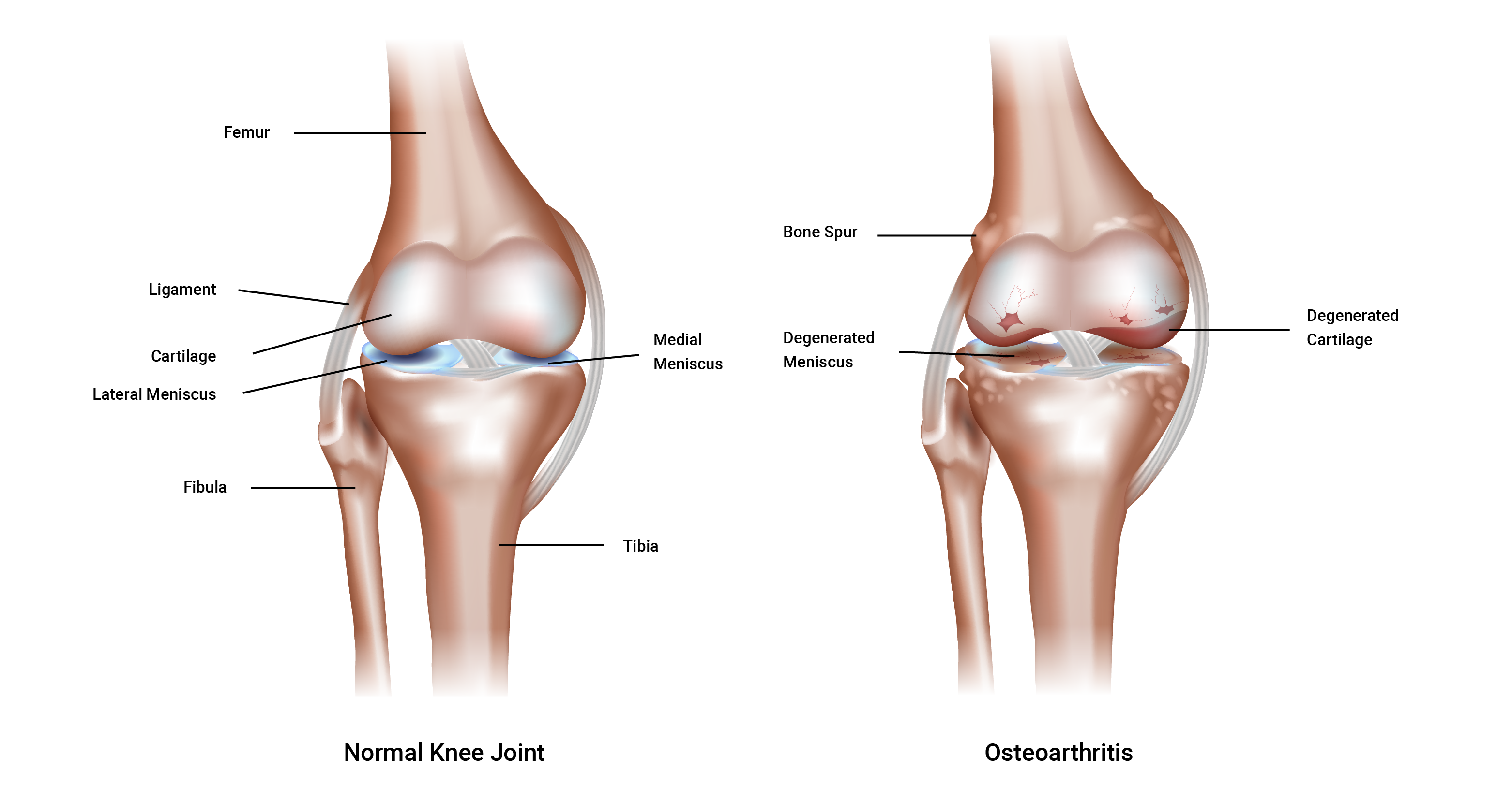
Osteoarthritis
Osteoarthritis is a largely age-related degeneration. Although this does not mean that younger generations are immune to osteoarthritis. It can be caused by wear and tear over time or disuse due to a lack of meaningful activity, leading to weakened (thinner) cartilage. This is referred to as a degenerative joint disease. The cartilage is lost from aging, obesity, joint irritation, muscle weakness, and general wear and tear. You can see the importance of effective exercise programming when working with clients who may have osteoarthritis.
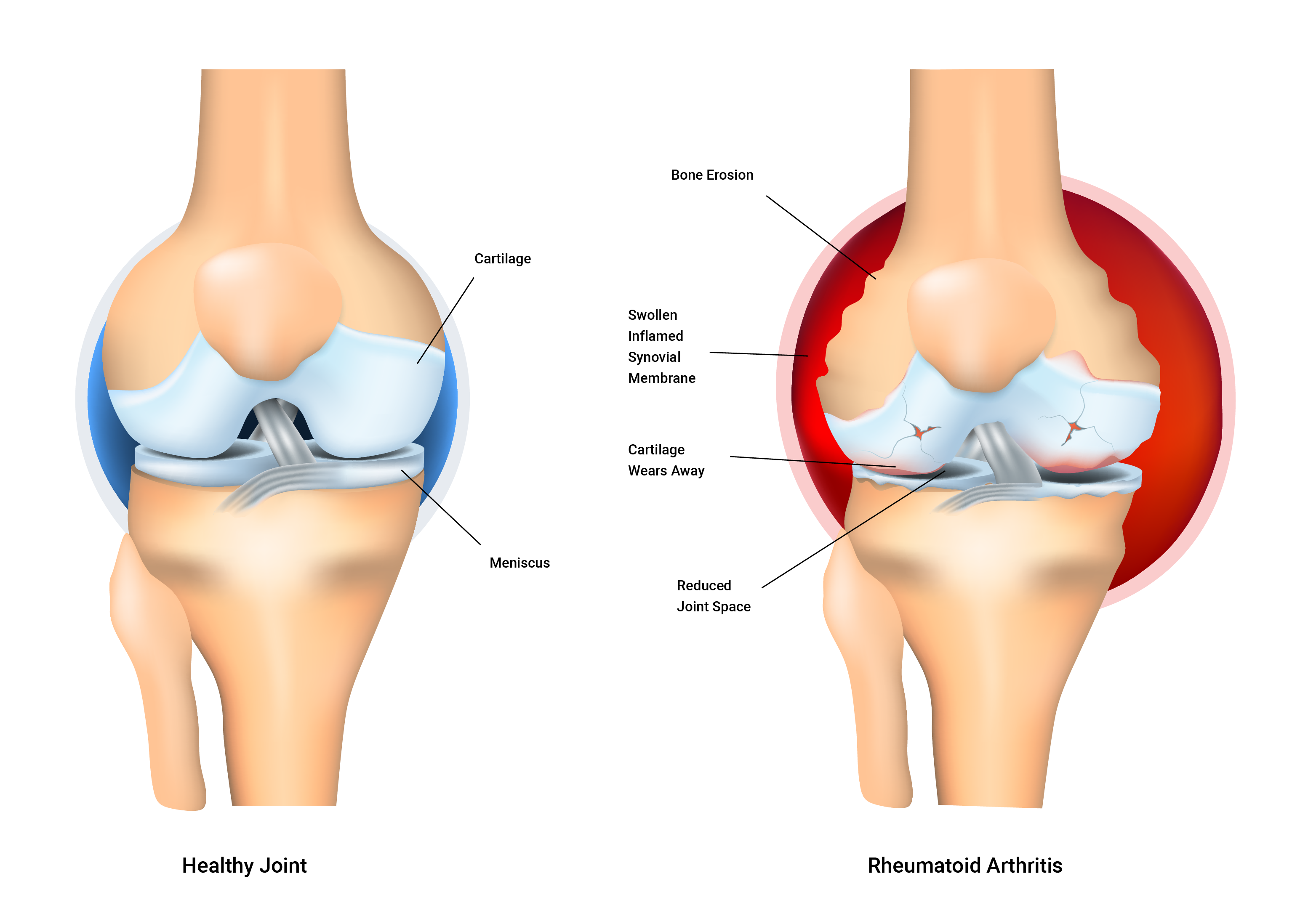
Rheumatoid arthritis
Rheumatoid arthritis is an auto-immune disorder. It occurs when your body’s immune system attacks the tissues of the body, in this case, its own joint linings and cartilage. These attacks affect the synovium.
Rheumatoid arthritis is a disease of the synovium that will invade and destroy a joint. It can eventually destroy the bone and cartilage inside the joint. It is typically characterised by inflammation of the joint, which can cause swelling, loss of function, and pain. Sadly, if one joint is affected, it is likely the other one will also be affected (but not usually to the same degree).
The exact cause of the immune system’s attacks is unknown. But scientists have discovered genetic markers that increase your risk of developing rheumatoid arthritis fivefold.
With exercise, the synovial fluid becomes less viscous and is more easily taken in by articular cartilage. This is beneficial because the synovial fluid provides essential nutrients to nourish the bone, and other cells remove unwanted debris to keep the synovial joint 'clean and tidy'. Cartilage acts like a sponge and has tiny pores that allow fluid to enter and leave.
Exercise design should consider the need for, and the benefit of, repetitive compression for nutrition and waste removal.
Connective tissue
There are 3 types of connective tissue within joints.
- Cartilage forms a cushion between bones to stop them from rubbing against each other.
- Ligaments are like strong cables or strings that hold bones together.
- Tendons attach muscles to bones or other muscles.
Joint stability vs range of motion
Unfortunately, it is difficult to achieve both ideal joint stability and ideal range of motion at the same time because an increase in one of these often occurs at the expense of the other. A balanced and well-thought-out programme design should keep this in mind.
Joint stability (resistance to displacement)
Joint stability is resistance to displacement and depends on 3 key factors:
- Joint ligaments
- Muscle tension
- Fascia tension
Let's look at each of these.
1. Ligaments
Ligaments are strong and flexible. They attach the ends of bones to form a movable joint. A ligament's main function is to maintain the correct relationship between bones. They also have receptors that monitor movement speed and range and resist undesirable movements.
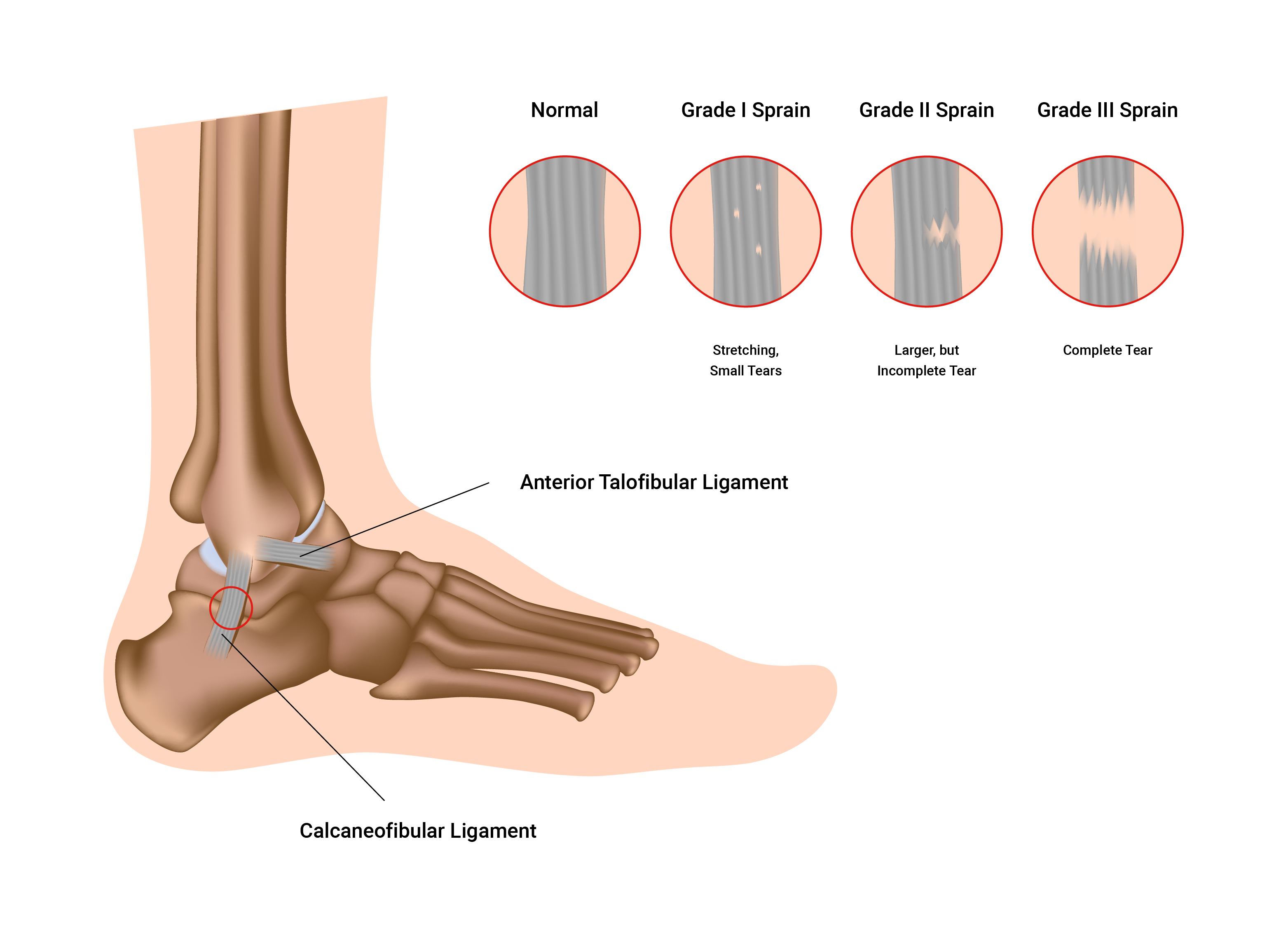
Ligament injury (sprain)
Ligaments are not very elastic. If they are over-stretched, they might never regain normal length.
Once stretched, their usefulness is permanently affected and joint stability is diminished. Think of an elastic band or a hair tie. Over time and repeated use, they stretch and begin to lose their shape (become bigger, and get looser). If left to dry, they become rigid and stiff.
Sprains range in severity, often referred to as a 'grade'. This grade outlines the current health and condition of the ligament(s) affected. Sprains range from first-degree to third-degree, with the third-degree often representing a severe (or full) rupture of the ligaments, as well as other symptoms. Recovery time also increases from first- to third-degree sprains. Typically ligaments (and tendons) have a lesser blood supply than muscle tissue which means that their nutrient delivery is less so recovery will be slower.
Think about the main function of the ligament - to attach the ends of bones to form a moveable joint and maintain the correct relationship between bones - as you study the following illustration.
2. Muscle tension
Muscles help static stability and provide dynamic stability. Strength training is an important defence against joint injury. Once ligaments are damaged (lengthened), muscles around the joint must be strengthened to maintain joint stability. This can lead to unfavourable changes in their range of motion.
3. Fascia tension
Fascia is a connective tissue that wraps around individual muscle fibres and groups of muscles. It is flexible and elastic within limits. This facia supports joint stability and integrity by supporting muscle attachment. It can also cross over joints to provide extra stability.
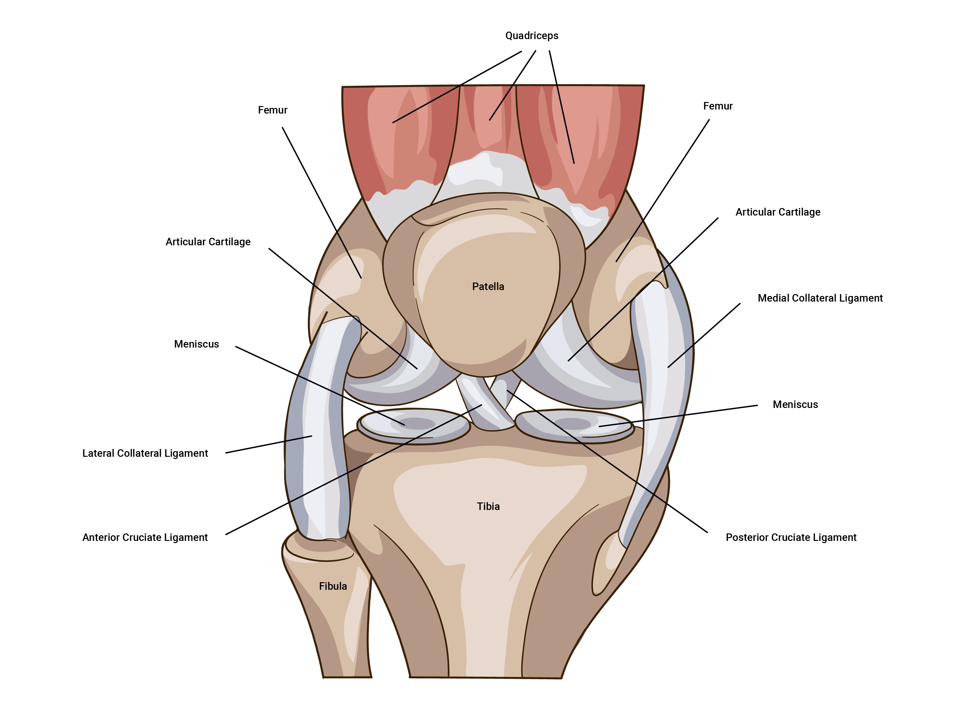
Menisci
Some loaded synovial joints (such as the knee) have flattened, shock-absorbing pads of fibrocartilage called menisci (the singular is meniscus) between the articulating surfaces of bones. These discs support the bones in the joint by binding strongly inside the joint capsule. They also offer depth to shallow joints and help absorb shock. The complete function of the menisci is not yet fully understood however they are known to be strong contributors to:
- absorbing shock.
- providing cushioning and padding in between bony surfaces.
- distributing synovial fluid over the articular surfaces of the joint.
Range of movement
3 key factors affect the range of joint movement.
- Muscles and their tendons (flexibility)
- Bulky tissue (fat, excessive muscle)
- Disease and/or condition (pain)
Flexibility of muscle and tendon
This is the single most important factor in joint range of movement. This can only be increased through dedicated and regular stretching. There is also a genetic predisposition to flexibility.
The bony structure of your joints will ultimately determine the range of motion you can achieve. I.e., deep vs shallow sockets, and the angle of the bones at insertion. Trying to stretch beyond your structural limitations will cause bone-on-bone contact and may result in bony lesions or spurs that result in ongoing pain.
Bulky tissue
Excess fat mass may make a person more restricted in their movement and function. It may also put additional strain and pressure on the internal organs. Excess muscle mass can restrict the range of movement at joints if flexibility is not maintained. It is possible to have both though!
Disease, illness, or condition
Joint disorders, such as arthritis and autoimmune diseases like lupus, can impact joints, making them stiff and swollen. Sprains and dislocations also cause restriction of the range of movement.
Before moving to the next topic, check that you have a good grasp of joints.
Watch
The following video provides a summary of joint types and classifications.