In this section you will learn to:
- Correctly use and interpret health terminology that describes the normal structure, function, and location of the major body systems
- Correctly use and interpret information that relates to the interrelationships between major components of each body system and other structures.
Supplementary materials relevant to this section:
- Reading A: Heimlich Manoeuvre
- Reading B: Imbalanced Breathing in Connection with Health Problem
- Reading C: Upper Respiratory Tract Infections
- Reading D: An Overview and Management of Osteoporosis
- Reading E: Full Body Stretching Routine
- Reading F: The Stroke Survivor
What is life? On one hand, this question seems easy and on the other hand so abstract, that it is more like a riddle. We all think we can recognise life even if we can’t define it easily.
Humans are composed of microscopic units known as cells, which are the ‘building blocks’ of life. Cells have a number of features in common, which allow them to grow, reproduce and generate more cells. Throughout this section of the Study Guide we will provide you with an overview of the human body focusing on the different structures, functioning and location of the major body systems. We will also look at specific body systems in detail and understand the relationship between the major components within these systems.

The basic building block of living organisms is the cell. The human body is composed of countless millions of cells. There are many different types of cells, which are specialised so they can carry out particular functions in the body. Despite these differences, certain features are the same in most cells. The figure below shows the structures present in a typical animal cell.

The living material that makes up a cell is called cytoplasm. It has a texture rather like sloppy jelly, somewhere between a solid and a liquid.
The largest organelle in the cell is the nucleus. Nearly all cells have a nucleus, with a few exceptions such as red blood cells. The nucleus controls the activities of the cell. It contains chromosomes (46 in human cells) which carry the genetic material or genes. Genes control the activities in the cell by determining which proteins the cell can make (see below). One very important group of proteins found in cells is enzymes. Enzymes control chemical reactions that take place in the cytoplasm.
All cells are surrounded by a cell membrane, sometimes called the cell surface membrane to distinguish it from other membranes inside the cell. This is a thin layer like a ‘skin’ on the surface of the cell. It forms a boundary between the cytoplasm of the cell and the outside.
However, it is not a complete barrier. In fact, as you will see, the membrane can actively control the movement of some substances.
One organelle that is found in the cytoplasm of nearly all living cells is the mitochondrion (plural mitochondria). In cells that need a lot of energy, such as muscle or nerve cells, there are many mitochondria. This gives us a clue as to their role. They perform some of the reactions of respiration, releasing energy that the cell can use. Most of the energy from respiration is released in the mitochondria.
Differentiation of cells
A human begins life as a zygote, which divides to form two cells, then four, then eight and so on, until an embryo is formed, containing many millions of cells.

As the developing embryo grows, cells become specialised to perform particular roles. This specialisation is also under the control of the genes and is called differentiation. Different kinds of cell develop depending on where they are located in the embryo, for example, a nerve cell in the spinal cord, or an epidermal cell in the outer layer of the skin.
Muscle Tissues

Muscle tissue consists of cells that are specialised to shorten, or contract, resulting in movement of some kind. Muscle tissue is composed of tightly packed cells called muscle fibres. The fibres are generally long and thin and aligned parallel to each other. Muscle fibre contains proteins which interact to make the cell contract. There are three types of muscle tissue: voluntary, involuntary and, cardiac. They vary somewhat in body location, structure, and function, but they all do essentially the same thing— when stimulated, they contract. For now, we’ll focus solely on differences between the three types of muscle tissue.
Skeletal muscle tissue (voluntary)
Skeletal muscle tissue connects to tendons, which attach to bones. When skeletal muscles contract, they cause body parts to move. A skeletal muscle may contain thousands of individual cells, all aligned parallel to each other. A skeletal muscle is called voluntary muscle because we can exert conscious control over its activity. Skeletal muscle cells are activated only by nerves.
Smooth muscle tissue (involuntary)
These tissues are hollow organs and tubes, including blood vessels, digestive tract, uterus, and bladder. These slim cells are much smaller than skeletal muscle cells and have only one nucleus, like cardiac muscle. The cells are aligned roughly parallel to each other. In blood vessels, they are generally aligned in a circular fashion around the vessel. When smooth muscle cells shorten, the diameter of the blood vessel is reduced. Smooth muscle cells taper at both ends, and there are gap junctions between adjacent cells so that when one contracts, nearby cells also contract. Like cardiac muscle, smooth muscle is involuntary in that we cannot control its contractions consciously
Cardiac muscle tissue
Cardiac muscle tissue (in Greek, kardia means “the heart”) is found only in the heart. The individual cells are much shorter than skeletal muscle fibres, and they have only one nucleus. Like skeletal muscle, the cells are arranged parallel to each other. Cardiac muscle cells are short and blunt ended, with gap junctions between the ends of adjoining cells. The gap junctions represent direct electrical connections between adjoining cells, so when one cell is activated, it activates it neighbours down the line. Because of these gap junctions, the entire heart contracts in a coordinated fashion. Cardiac muscle is considered involuntary because the heart can contract rhythmically entirely on its own, without any conscious thought on our part and without any stimulation by nerves.
TIP
Skeletal muscle tissue connects to tendons and bones. Smooth muscle tissue surrounds organs and tubes. Cardiac muscle tissue comprises most of the heart. The common feature of all muscle tissues is that they contract, producing movement
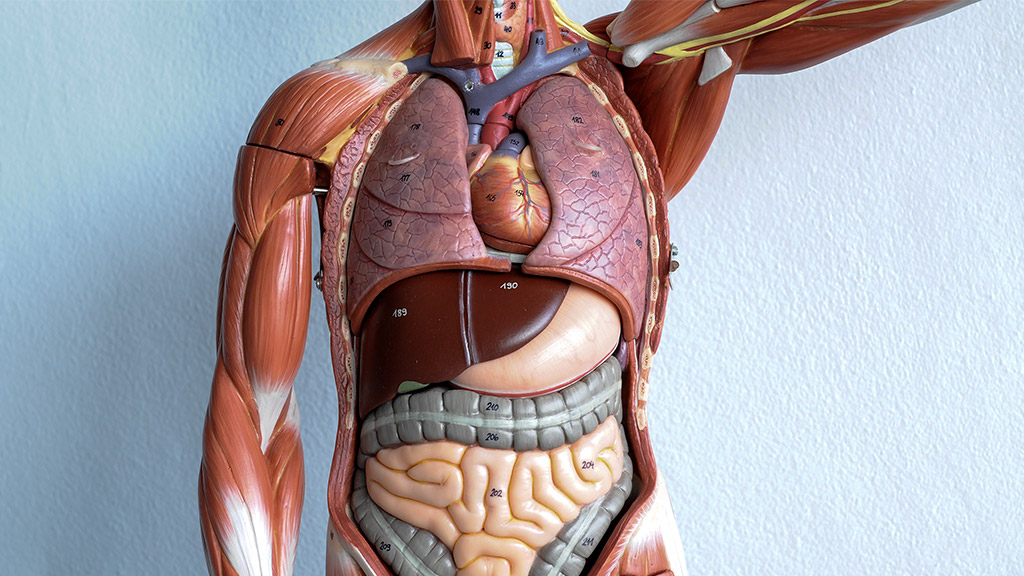
Other tissues include blood, which is made of various types of red and white blood cells in a liquid matrix called plasma, and nervous tissue, which makes up the brain, spinal cord, and nerves. A collection of different tissues carrying out a particular function is called an organ. The main organs of the human body are shown in the image below.


The human body is a very complex machine. It needs food for growth and repair of tissues, as well as for a supply of energy. In this section, we look at how the cells and tissues we spoke about above are organised into organs and organ systems that comprise your body, as well as how they work together to maintain the health and stability of your body.

Many of the more complex functions of multicellular organisms (such as pumping blood or digesting food) cannot be carried out by one tissue type alone. Organs are structures composed of two or more tissue types joined together that perform a specific function or functions. Your heart is an organ because the tissues that comprise it share one essential function: They pump blood. Most of the heart consists of cardiac muscle, but there is also smooth muscle in the blood vessels that supply the cardiac muscle. The heart contains nervous tissue that affects the rate at which the organ beats. Some connective tissue, primarily in the heart valves, open and close to control blood flow within the heart, and even a thin layer of epithelial tissue lines the heart chambers. Some organs have several functions. For example, the kidneys remove waste and helps control blood pressure.
Organ systems are groups of organs that together serve a broad function that is important to survival either of the individual organism or a species. For example, survival of an individual organism depends on respiration, movement, or excretion of wastes, and survival of a species relies on reproduction. A good example is the organ system responsible for the digestion of food. Your digestive system includes your mouth, throat, esophagus, stomach, intestines, and even your liver, pancreas, and gallbladder. All these organs must interact and be controlled and coordinated to accomplish their overall function.
(Johnson, 2017)

Cells get their energy by oxidising foods such as glucose. This process is called respiration and needs a continuous supply of oxygen from the blood. In addition, the waste product carbon dioxide needs to be removed from the body. These gases are exchanged between the blood and the air in the lungs. In this section, you will learn about the processes of respiration and gas exchange.
Tip
Respiration is called an oxidation reaction because oxygen is used to break down food molecules.
The organs of the respiratory system, which are located in the chest cavity, ensure that oxygen enters the body and carbon dioxide leaves the body. During inspiration, or inhalation (breathing in), air is conducted from the atmosphere to the lungs by a series of cavities, tubes, and openings. During expiration, or exhalation (breathing out), air is conducted from the lungs to the atmosphere by way of the same structures.
The respiratory tract extends from the nose to the lungs. Note the organs in the upper respiratory tract and the ones in the lower respiratory tract.

The upper respiratory tract
The nasal cavities, pharynx, and larynx are the organs of the upper respiratory tract.
The nose
The nose opens at the nares (nostrils) that lead to the nasal cavities. The nasal cavities are narrow canals separated from each other by a septum composed of bone and cartilage. Air entering the nasal cavities encounters large, stiff hairs that act as a screening mechanism. The hairs filter the air and trap small particles (dust, mould spores, pollen, etc.) so that they don’t enter air passages. The rest of the nasal cavities are lined by mucous membrane.

Did you know that?
Scientists have found that air travels at 100 miles an hour during a sneeze. This is enough force to propel sneeze droplets up to 12 ft away from the person sneezing. If a sneeze is stifled, the air is forced into the auditory (eustachian) tube and middle ear, potentially causing damage to the middle ear.
The pharynx
The pharynx, commonly referred to as the throat, is a funnel shaped passageway that connects the nasal and oral cavities to the larynx. The pharynx has three parts: the nasopharynx, where the nasal cavities open above the soft palate; the oropharynx, where the oral cavity opens; and the laryngopharynx, which opens into the larynx.
The larynx
The larynx is normally open, allowing air to pass, but the oesophagus is normally closed and opens only when a person swallows. If someone swallows and some of the food enters the larynx, coughing occurs in an effort to dislodge the food. If the passageway remains blocked, the Heimlich manoeuvre can be used to dislodge food blocking the airway.
Reading A: Heimlich Manoeuvre
Reading A explains the steps taken in a Heimlich Manoeuvre, which is a rapid first-aid procedure to treat choking due to upper airway obstruction by a foreign object.
When a person’s air supply is partly or fully blocked, they are at risk of injury and death from choking or suffocation. This can occur when breathing is disrupted by a physical blockage, such as an object in the windpipe, or when a person is trapped in a low oxygen environment.
Choking and suffocation injuries resulted in 1700 hospitalisations between 2020 and 2021, which is 6.5% per 100,000 population.
This represents 7% of injury deaths and 0.3% of injury hospitalisations.
Choking on objects is the most common cause of death in this category, while a foreign body in the respiratory tract is the most common cause of hospitalised injury.
Older people have the highest rates of death by choking and suffocation, while young children have the highest rates of hospitalisation.
Rates of death due to choking and suffocation were highest for people aged sixty-five and over, while young children aged 0 to 4 had the highest rates of hospitalisation compared with other life-stage age groups.
Of the hospitalisations in 2020–21:
Thirty-two percent were for people aged 65 and over
26% were for children aged 0–4.
Males were 1.3 times as likely as females to be hospitalised for choking or suffocation in 2020–21.
Of the deaths in 2019–20, 80% were for people aged 65 and over.

(Australian Institute of Health and Welfare, November 2022)
The lower respiratory tract

Once the incoming air makes its way past the larynx, it enters the lower respiratory tract. The lower respiratory tract consists of the trachea, the bronchial tree, and the lungs.

The trachea

The trachea, commonly called the windpipe, is a tube connecting the larynx to the primary bronchi. It is separated from the oesophagus by a flexible, muscular wall. This orientation allows the oesophagus to expand when swallowing. The goblet cells produce mucus, which traps debris in the air as it passes through the trachea. The mucus is then swept toward the pharynx and away from the lungs by the cilia that project from the epithelium. When you cough, the tracheal wall contracts, narrowing its diameter.
Therefore, coughing causes air to move more rapidly through the trachea, helping expel mucus and foreign objects. Smoking is known to destroy the cilia; consequently, the soot in cigarette smoke collects in the lungs. Smokers often develop heavy coughs as a result. If the trachea is blocked because of illness or the accidental swallowing of a foreign object, a breathing tube can be inserted by way of an incision made in the trachea. This tube acts as an artificial air intake and exhaust duct. The operation is called a tracheostomy.
Tobacco smoking is one of the largest preventable causes of death and disease in Australia. Smoking is estimated to kill almost 20,500 Australians a year (13% of all deaths) and was responsible for 8.6% of the total burden of disease in Australia in 2018. It is associated with an increased risk of health conditions including heart disease, diabetes, stroke, cancer, kidney disease, eye disease and respiratory conditions such as asthma, emphysema, and bronchitis.
In 2020-21, one in ten people (10.7% or 2.1 million people) aged 18 years and over were current daily smokers, with men more likely than women to smoke daily (12.6% compared to 8.8%). One in twelve (8.3%) people aged 18-24 years smoked daily, and this increased with age until 55-64 years where the rate peaked at 13.7%.

(Australian Bureau of Statistics, March 2022)
The bronchial tree (bronchi/bronchioles)

The trachea divides into right and left primary bronchi (singular term - bronchus), which lead into the right and left lungs. The bronchi branch into a few secondary bronchi that also branch, until the branches become bronchioles, which are about 1 mm in diameter. The bronchi resemble the trachea in structure. As the bronchial tubes divide and subdivide, their walls become thinner, and the small rings of cartilage are no longer present.
During an asthma attack, the smooth muscle of the bronchioles contracts, causing bronchiolar constriction and characteristic wheezing. Each bronchiole leads to an elongated space enclosed by a multitude of air pockets, or sacs, called alveoli.
Asthma is a long-term lung condition that is caused by narrowing of the airways when they become inflamed [1]. People with asthma have trouble breathing, and the most common symptoms are wheezing, coughing, breathlessness, and chest tightness. Asthma may affect people of all ages and can usually be managed through treatment such as medication use (e.g., reliever inhalers and preventer/controller medications) and a healthy lifestyle.
Just under 2.7 million people (10.7% of the total population) had asthma in 2020-21. Females were more likely than males to have asthma (12.0% compared to 9.4%); however, the rate of asthma was similar in children aged 0-14 years (9.5% and 7.9%).

(Australian Bureau of Statistics, July 2021)
The lungs
The lungs are paired, cone-shaped organs in the thoracic cavity. In the centre of the thoracic cavity are the trachea, heart, thymus, and oesophagus. The lungs are on each side of the trachea. The right lung has three lobes, and the left lung has two lobes, allowing room for the heart, which points left. Each lobe is further divided into lobules, and each lobule has a bronchiole serving many alveoli.
Pneumonia is a common and potentially fatal lung infection that should not be underestimated. It can be caused by viruses, bacteria, or fungi. When you breathe, air travels through the lungs to the alveoli or air sacs. If you have pneumonia the air sacs in your lungs fill with secretions and fluids that obstruct normal air flow. There are many types of pneumonia, one of the most common and life-threatening types is pneumococcal pneumonia, which is caused by the bacterium, Streptococcus pneumoniae.
This bacterium is responsible for approximately 1.6 million deaths each year world-wide. Healthy people may carry it in their nose and throat. While most of the time this does not cause any illness, vulnerable groups such as those over the age of 70, infants and people with a chronic illness may develop pneumococcal disease.
(Lung Foundation Australia, n.d.)
Tip
The thin layer of fluid lining the inside of the alveoli comes from the blood. The capillaries and cells of the alveolar wall are ‘leaky’, and the blood pressure pushes fluid out from the blood plasma into the alveolus. Oxygen dissolves in this moist surface before it passes through the alveolar wall into the blood. If you want to know more about pneumonia clicks on the link - https://www.youtube.com/watch?v=JxZBbuaPxJk&t=25s
Mechanism of Breathing (The basics of breathing in and out)

Ventilation means moving air in and out of the lungs. This requires a difference in air pressure – the air moves from a place where the pressure is high to one where it is low. Ventilation depends on the fact that the thorax is an airtight cavity. When we breathe, we change the volume of our thorax, which alters the pressure inside it. This causes air to move in or out of the lungs. There are two movements that bring about ventilation, those of the ribs and the diaphragm.
If you put your hands on your chest and breathe in deeply, you can feel your ribs move upwards and outwards. They are moved by the intercostal muscles. The outer (external) intercostals contract, pulling the ribs up. At the same time, the muscles of the diaphragm contract, pulling the diaphragm down into a more flattened shape. Both these movements increase the volume of the chest cavity and cause a slight drop in pressure inside the thorax compared with the air pressure outside. Air then enters the lungs (inhalation or inspiration).

The opposite happens when you breathe out deeply. The external intercostals relax and the internal intercostals contract, pulling the ribs down and in. At the same time, the diaphragm muscles relax, and the diaphragm goes back to its normal dome shape. The volume of the thorax decreases and the pressure in the thorax is raised slightly above atmospheric pressure. This time, the difference in pressure forces air out of the lungs. Exhalation (expiration) is helped by the fact that the lungs are elastic, so they tend to collapse and empty like a balloon.
Changes in the position of the ribs and diaphragm during breathing.
- breathing in (inhalation)
- breathing out (exhalation)
It is important that you remember the changes in volume and pressure during ventilation. If you have trouble understanding them, think of what happens when you use a bicycle pump. If you push the pump handle, the air in the pump is squashed, its pressure rises, and it is forced out of the pump. If you pull on the handle, the air pressure inside the pump falls a little and air is drawn in from outside. This is similar to what happens in the lungs.
Reading B: Imbalanced Breathing in Connection with Health Problems
Reading B describe the most common direct or indirect connections between imbalanced breathing and health. Imbalanced breathing potentiates symptoms, thus perpetuating the problematic situation. This is very common in states of overarousal, such as stress, anxiety, and pain. In addition, through stress reactions, conditions such as physical illness or surgery may contribute to the development of imbalanced breathing patterns and aggravation of symptoms.
Gas exchange

You can tell what is happening during gas exchange if you compare the amounts of different gases in atmospheric (inhaled) air with the air breathed out. The table below shows the approximate percentage volume of gases in inhaled (atmospheric) and exhaled air. Exhaled air is also warmer than inhaled (atmospheric) air and is saturated with water vapour.
| Gas | Inhaled (atmospheric) air | Exhaled air |
|---|---|---|
| Nitrogen | 78 | 79 |
| Oxygen | 21 | 16 |
| Carbon dioxide | 0.04 | 4 |
| Other gases | 1 | 1 |
Reading C: Upper Respiratory Tract Infection
Reading C examines when an upper respiratory tract infection should be considered on differential diagnosis and how to properly evaluate it.

Humans are vertebrates – the human body is supported by an internal skeleton made of bone, which has a central ‘axis’ comprising the vertebral column, or backbone. Attached to the bones of the skeleton are voluntary muscles. These muscles are stimulated to contract by the nervous system, under conscious control by the brain. Contraction of the muscles pulls on bones, which produces movement.

The Skeleton
The human skeleton has several functions. It protects many important organs. For example, the cranium (skull) protects the brain, eyes and ears, the vertebrae protect the spinal cord, and the ribcage protects the heart and lungs. Bones also make some components of blood. Red blood cells and platelets are made in the bone marrow of larger bones, such as the sternum, femur, and pelvis. The skeleton’s most obvious roles, however, are in support and movement.
Tip
Osteoporosis is a medical condition that affects elderly people. Their bones lose calcium salts and become porous and less dense, so that they break easily. Women are particularly vulnerable after the menopause, due to changes in hormone levels. There is no cure for osteoporosis, but it can be helped by good diet, calcium, and vitamin D supplements.
When humans move, their bones move in relation to each other. The point where two bones meet is called a joint. We say that the bones articulate at joints. A movable joint, such as the hip or elbow, needs to have certain features. These include:
- a way to keep the ends of the bones together, so that they do not separate (dislocate)
- a means of reducing friction between the ends of the moving bones
- a shock-absorbing surface between the two bones.
There are various kinds of joint in the body. They are classified into three groups, according to the amount of movement they allow:
- freely movable joints/ball and socket – hip joint, knee, and wrist
- partially movable joints - vertebrae
- immovable (fixed) joints – skull
Freely movable joints include ball and socket joints, such as the shoulder and hip, and hinge joints, such as the elbow.
Tip
A ball and socket joint allows movement in any direction. Imagine your shoulder joint is at the corner of a box. You can move your upper arm in any of the three directions formed by the sides of the box: side, back or top. Compare this with a hinge joint, such as your elbow – due to the shape of the joint, you can only move this in one direction, as when bending your arm.

In summary, the skeleton does more than merely provide a frame for the body. In addition, it has the following functions:
- Support - The bones of the legs support the entire body when we are standing, and bones of the pelvic girdle support the abdominal cavity.
- Movement - The skeletal system works with the muscular system to provide movement.
- Protection - The bones of the skull protect the brain; the rib cage protects the heart and lungs; and the vertebrae protect the spinal cord, which makes nervous connections to all the muscles of the limbs.
- Production of blood cells - The skeleton plays an important role in the formation of blood. In the foetus, all bones in the foetus have red bone marrow to produce new blood cells. However, in adults, only certain bones produce blood cells.
Reading D: An overview and management of osteoporosis
Reading D looks at how osteoporosis is related to various factors such as menopause and aging and provides a detailed explanation of this chronic metabolic bone disease
Muscles
Skeletal muscles are organs that are attached to bones and move by contracting (becoming shorter), pulling on the bone. At the end of a muscle, there are tendons. A tendon attaches the muscle to the bone. Tendons have very high tensile strength, like ligaments, but unlike ligaments they are not very elastic. This means that they do not stretch when the muscle contracts. Muscles cannot push, they can only pull – in other words, they are not able to expand actively.
In 2017-18, 924,000 Australians (3.8%) had osteoporosis. This was like 2014-15 (3.5%). Like arthritis, osteoporosis was more common amongst females than males (6.2% prevalence compared with 1.5%), and more common in older age groups. The increase in older age groups was more apparent and began earlier in females than in males. For females, there was an increase in the proportion who had osteoporosis starting at the 45–54-year age group, with continuous increases with each successive age group. The first increase for males, in contrast, was in the 55–64-year age group, with no further increases until 75 years and over. Females aged 75 years and over were almost three times more likely than males to have osteoporosis (29.0% compared with 10.3%).
(Australian Bureau of Statistics, December 2018)
Reading E: Full Body Stretching Routine
Reading E takes a closer look at the numerous benefits of full body stretching and how to build a stretching routine that targets all your major muscle groups.

The nervous system is a coordination system forming a link between stimulus and response.

The body has a second coordination system, which does not involve nerves. This is the endocrine system. It consists of organs called endocrine glands, which make chemical messenger substances called hormones. Hormones are carried in the bloodstream.

The main endocrine glands, including the hormones they make and related functions, are shown in the table below:
| Gland | Hormone | Main Functions |
|---|---|---|
| Pituitary | Follicle stimulating hormone (FSH) | Stimulates egg development and oestrogen secretion in females and sperm production in males |
| Growth hormone | Speeds up the rate of growth and development in children | |
| Luteinising hormone (LH) | Stimulates egg release (ovulation) in females and testosterone production in males | |
| Thyroid | Thyroxine | Controls the body’s metabolic rate (how fast chemical reactions take place in cells) |
| Pancreas | Insulin | Lowers blood glucose |
| Glucagon | Raises blood glucose | |
| Adrenals | Adrenaline | Prepares the body for physical activity |
| Testes | Adrenaline | Controls the development of male secondary sexual characteristics |
| Ovaries | Oestrogen | Controls the development of female secondary sexual characteristics |
| Progesterone | Regulates the menstrual cycle |
Self-Reflection
Many hormones participate in negative feedback loops that maintain homeostasis. Steroid hormones enter the target cell, activate specific genes, and cause the production of new proteins. Nonsteroid hormones bind to a cell membrane receptor that either opens or closes ion channels or activates a second messenger within the cell.
Let’s now turn to some of the key endocrine glands and their hormones.
The hypothalamus
It is a small region in the brain that serves as a homeostatic control centre. It is an important link between the nervous system and the endocrine system. The hypothalamus receives neural input about certain internal environmental conditions such as water and solute balance, temperature, and carbohydrate metabolism. It produces
The thyroid and parathyroid
These glands are anatomically linked. The thyroid gland is situated just below the larynx at the front of the trachea, and the two lobes of the thyroid gland wrap part of the way around the trachea. The thyroid and parathyroid glands are also functionally linked; both help to regulate calcium balance. In addition, the thyroid has a separate and important role in controlling metabolism. The two main hormones produced by the thyroid gland are thyroxine and calcitonin. The parathyroid glands produce parathyroid hormone (PTH), which increases the absorption of calcium by the digestive tract and causes the kidneys to retain calcium and excrete phosphate.

The pituitary gland
It is found at the base of the brain. It secretes a number of hormones, including several that regulate reproduction. It is sometimes called the “master gland” because it secretes eight different hormones that in turn regulated many of the other endocrine glands. It consists of two lobes:
- Posterior lobe - is the back lobe of your pituitary gland, which is a small, pea-sized gland located at the base of your brain below your hypothalamus. The posterior pituitary stores and releases just two of the many hormones your pituitary gland is responsible for: oxytocin and antidiuretic hormone (ADH, or vasopressin).
- Anterior lobe - is the front lobe of your pituitary gland, which is a small, pea-sized gland located at the base of your brain below your hypothalamus. It is responsible for 6 different hormones – growth, reproduction, metabolism, stress and lactation.
The pancreas
It is both an endocrine and an exocrine organ. It secretes two hormones called insulin and glucagon, both of which are involved in the regulation of blood glucose. The pancreas is also a gland of the digestive system, secreting enzymes through the pancreatic duct into the small intestine.
The pineal glands
The name pineal derives from the fact that the gland is shaped like a small pinecone (pineas in Latin). The pineal secretes the hormone melatonin (not to be confused with the skin pigment, melanin) in a cyclic manner, coupled to the daily cycle of light and dark. The pineal gland is a pea-sized gland located deep within the brain.
The human gonads
These are the testes for makes and the ovaries for the females.
- Testes produce testosterone
The testes, located in the scrotum, produce androgens, the male sex hormones. The primary androgen produced by males is testosterone. In males, testosterone regulates the development and normal functioning of sperm, the male reproductive organs, and male sex drive. It is also responsible for the spurt of bone and muscle growth at puberty and the development of male secondary sex characteristics such as body hair and a deepening voice - Ovaries produce estrogen and progesterone
The ovaries, located in the abdomen, produce the female sex hormones known collectively as estrogens. Estrogen initiates the development of female secondary sex characteristics such as breast development, widening pelvis, and distribution of body fat. Both estrogen and progesterone regulate the menstrual cycle, the monthly changes in the tissue of the uterus that prepare the female body for pregnancy.

The biological name for a nerve cell is a neurone. The impulses that travel along a neurone are caused by movements of charged particles (ions) in and out of the neurone. Impulses travel at speeds between about 10 and 100 metres per second.
The nervous system receives information from many different senses simultaneously, and then integrates this information. This means it acts very fast. It can receive information, integrate it, and produce a response within tenths of a second. The nervous system can also initiate specific responses, including muscle contraction, glandular secretion, and even conscious thought and emotions.
Most of the functions of the nervous system are completely automatic and can be carried on simultaneously without requiring our attention or conscious decision making. Working with the endocrine system, the nervous system maintains homeostasis. It also allows us to feel emotions, to be aware of ourselves, and to exert conscious control over the incredible diversity of our physical movements.
Neurons

Neurons are cells specialised for communication. They generate electrical impulses from one of part of the body to another. The longest neurons extend all the way from your toes to your spinal cord. There are three types of neurons:
- Sensory neurons – responds to certain stimulus such as light or pressure
- Motor neurons – carries the nervous system’s output to all of the tissues and organs of the body.
- Interneurons – these neurons receive input from a sensory neuron, integrate this information and influence the functioning of other neurons.
The brain
Your brain is sometimes called your ‘grey matter’. This is because the positions of the grey and white matter are reversed in the brain compared with the spinal cord. The grey matter, mainly made of nerve cell bodies, is on the outside of the brain, while the axons that form the white matter are in the middle of the brain. The brain is made up of different parts, each with a specific function.

The largest part of the brain is the cerebrum, made of two cerebral hemispheres. The cerebrum is the source of all our conscious thoughts. It has an outer layer called the cerebral cortex, with many folds all over its surface. The cerebrum has three main functions.
- It contains sensory areas that receive and process information from all our sense organs.
- It has motor areas, from which all our voluntary actions originate.
- It is the origin of ‘higher’ activities, such as memory, reasoning, emotions, and personality
The spinal cord
The spinal cord provides a means of communication between the brain and the peripheral nerves that leave the cord. When someone touches your hand, sensory receptors generate nerve signals that pass through sensory fibres to the spinal cord and up ascending tracts to the brain.

The brain coordinates the voluntary control of our limbs. Motor signals originating in the brain pass down descending tracts to the spinal cord and out to our muscles by way of motor fibres. Therefore, if the spinal cord is severed, we suffer a loss of sensation and a loss of voluntary control— paralysis. If the cut occurs in the thoracic region, the lower body and legs are paralysed, a condition known as paraplegia. If the injury is in the neck region, all four limbs are usually affected, a condition called quadriplegia.
Tip
Why does a stroke on the right side of the brain cause weakness or paralysis on the left side of the body?
- Motor neurons in the right cerebral hemisphere control the left side of the body and vice versa because of crossing-over. Likewise, sensation from the left half of the body travels to the right cerebral hemisphere. Destruction of brain tissue by a stroke interferes with outgoing motor signals to the opposite side of the body, as well as incoming sensory information from that side.
Reading I: The Stroke Survivor
Reading I is derived from the story of a stroke survivor, the individuals explain the onset of a stroke and the aftereffects of a stroke together with its prognosis.

The surroundings outside the body are called the external environment. The inside of the body is known as the internal environment. We are usually faced with a stimulus and a response. A stimulus is a change in a person’s environment and a response is a reaction to that change.
The Eye

The tough outer coat of the eye is called the sclera. This is the visible, white part of the eye. At the front of the eye, the sclera becomes a transparent ‘window’ called the cornea, which lets light into the eye.
Behind the cornea is the coloured ring of tissue called the iris. In the middle of the iris is a hole called the pupil, which lets the light through. It is black because there is no light escaping from the inside of the eye.
The innermost layer of the back of the eye is the retina. This is the light sensitive layer, the place where light energy is converted into the electrical energy of nerve impulses. The retina contains cells called rods and cones. These cells react to light, producing impulses in sensory neurones. The sensory neurones then pass the impulses to the brain through the optic nerve.
Self-Reflection
A way to prove to yourself that the eyes form two overlapping images is to try the ‘sausage test’. Focus your eyes on a distant object. Place your two index fingers tip to tip, and bring them up in front of your eyes, about 30 cm from your face, while still focusing on a distance. You should see a finger ‘sausage’ between the two fingers. Now try this with one eye closed. What is the difference?
The Ear

The ear has three parts: the outer, middle, and inner ear. The outer ear directs sound waves to the eardrum at the end of the auditory canal, causing it to vibrate. The vibrations are passed across the middle ear by three small bones (ear ossicles), the malleus (hammer), incus (anvil) and stapes (stirrup). These bones amplify the vibrations as they pass across them. The stapes transmits the vibrations to a membrane-covered opening called the oval window, which is at one end of a coiled structure called the cochlea.
The Eustachian tube connects the middle ear with the throat and allows the air pressure to be equalised either side of the eardrum
DID YOU KNOW?
Why do your ears ‘pop’ in an aeroplane? As an aeroplane gains height, the air pressure falls, causing the eardrum to bulge outwards. If you swallow, the Eustachian tube opens and allows air to pass into the middle ear from the throat, equalising the pressure. The ‘pop’ is caused by the eardrum going back to its normal position.

The sensations provided by receptors are categorised as either somatic or special.
- Somatic sensations are those that originate from receptors present at more than one location in the body.
- The five special senses originate from receptors that are restricted to particular areas of the body such as the ears and eyes.
Smell

Odours are detected by olfactory receptor cells located in the upper part of the nasal passage and causes them to generate ‘good or bad’ smell impulses.
Our sense of smell complements our sense of taste because when food is chewed, it releases chemicals that come into contact with olfactory receptors.
When we have a cold, most food taste less appetising, this is because we produce more mucus which prevents the odorant molecules from reaching the olfactory receptors impairing our sense of smell.
Taste

The tongue has a rough texture because its surface is composed of numerous small projections, called papillae, surrounded by deep folds. The exposed tip of a taste cells has hairs that project into the mouth.
The hairs contain chemoreceptors that are specific for certain chemicals called tastants. The five commonly accepted primary taste qualities are sweet, sour, salty and bitter. Most of the taste receptors are located around the edges, front and back of the tongue. The tongue is slightly more sensitive to sweet tastes on the tip, sour on the sides, salty on the top and sides and bitter towards the back.
Hearing

Since the parts and functions of the ear were covered in the previous section, we will have a look at how sound is transmitted to the ear. Sounds are waves of compressed air. Sound intensity is a physical property of sound that refers to the energy of the sound, whereas loudness refers to our subjective interpretation of sound. Both intensity and perceived loudness are related to the amplitude of the sound waves.
When waves of sound strike the ear, they cause vibration-sensitive mechanoreceptors deep within the ear to vibrate. The vibration causes the production of impulses that travel to the brain for interpretation.
| Sound | Decibels | How it affects your hearing |
|---|---|---|
| Rustling Leaves | 20 | Does not cause hearing loss |
| Normal conversation | 50-60 | Does not cause hearing loss |
| Vacuum cleaner | 70 | Does not cause hearing loss |
| Power lawn mower | 85-100 | Cumulative exposure causes hearing loss; newer mowers may be quieter |
| MP3 player max, ear buds | 105-120 | Risk of permanent hearing loss |
| Shotgun | 140 | Repeated exposure likely to cause permanent hearing loss |
| Jet engines at 100ft | 150 | Exposure likely to cause permanent hearing loss |
Equilibrium

Along with hearing, the inner ear is responsible for encoding information about equilibrium, the sense of balance. Each of the semicircular canals end in a space that has small hair cells in it. These spaces are called ampullae - whenever we turn our head, the inner ear turns along with it.
Each of the three semicircular canals is responsible for a specific direction of head movement: One of the canals responds to the head
- tilting upwards or downwards,
- one responds to it tilting to the right or to the left, and
- one responds to it turning sideways.

Take a look at the following image of the human digestive system. It is simplified so that you can see the order of the organs along the gut. The real gut is much longer than this and coiled up so that it fills the whole space of the abdomen.

Overall, its length in an adult is about 8 m. This gives plenty of time for the food to be broken down and absorbed as it passes through the gut. Food is broken down in the mouth, in the stomach and in the first part of the small intestine (called the duodenum). The food is broken down using enzymes, which are made in the wall of the gut or by glands such as the salivary glands and the pancreas.
Digestion begins in the mouth. Saliva helps moisten the food and contains the enzyme amylase, which starts the breakdown of starch. The chewed lump of food, mixed with saliva, then passes along the oesophagus (gullet) to the stomach. The food is held in the stomach for several hours, while initial digestion of protein takes place. The stomach wall secretes hydrochloric acid, so the stomach contents are strongly acidic. This has a very important function. It kills bacteria that are taken into the gut along with the food, helping to protect us from food poisoning.
Digestion continues in the last part of the small intestine (the ileum), and it is here that the digested food is absorbed. The last part of the gut, the large intestine, is mainly concerned with absorbing water out of the remains and storing the waste products (faeces) before they are removed from the body.

Reproduction allows humans to pass on their genes. This happens at fertilisation, through the fusion of special sex cells or gametes – the sperm and egg cells. In this section, you will learn about human reproduction, from production of gametes through to the birth of the baby, as well as growth and development to adulthood.
Humans reproduce sexually, producing specialised sex cells called gametes. The male gamete is a mobile cell called a sperm. The female gamete is a stationary egg cell (also called an ovum). The sperm must move to the egg and fuse (join) with it. This is called fertilisation.
There are four main stages in sexual reproduction.
- Gametes (sperm and eggs) are produced.
- The male gamete is transferred to the female gamete.
- Fertilisation occurs – the sperm fuses with the egg.
- A zygote is formed, which develops into a new individual
Unlike many other animals, humans are not reproductively capable at birth. Instead, they undergo a sequence of events called puberty, during which a child becomes a sexually competent young adult. The reproductive system does not begin to fully function until puberty is complete. Sexual maturity typically occurs between the ages of 10 and 14 in girls and 12 and 16 in boys. At the completion of puberty, the individual is capable of producing children.
The female reproductive systems
The female gonads are paired ovaries that produce eggs, also called ova, and the female sex hormones estrogen and progesterone. The uterine tubes conduct eggs and is also the main location of where fertilisation takes place. The uterus on the other hand, houses the developing foetus. The cervix contains opening to uterus which houses a developing foetus. The vagina receives the penis during intercourse and also serves as a birth canal and an exit for menstrual flow.

The menstrual cycle
Menstrual’ means ‘monthly’, and in most women the menstrual cycle takes about a month, although it can take as little as two weeks or as long as six weeks. In the middle of the cycle is an event called ovulation, which is the release of a mature egg cell (ovum). A cycle is a continuous process, so it does not really have a beginning, but the first day of menstruation is usually called day 1.

- 1-7 Days: Uterous lining breaks down, menstruation occurs.
- 8-11 Days: The lining of the womb thickens in preparation for the egg.
- 12-17 Days: Ovulation Occurs (Usually on day 14).
- 18-25 Days: If fertilization has not taken place, the corpus luteum fades away.
- 26-28 Days: The uterine lining detaches leading to menstruation.
Fertilisation
Each month, an egg is released into an oviduct from one of the ovaries. This is called ovulation. If an egg is present in the oviduct, it may be fertilised by sperm introduced during intercourse. The zygote formed begins to develop into an embryo, which sinks into (‘implants’ in) the lining of the uterus. Here, the embryo develops an organ called the placenta, which allows it to exchange substances with the mother’s blood. The placenta also maintains the embryo’s position in the uterus.
Foetus development
Human development proceeds from pre-embryonic to embryonic development and then through foetal development.
| Time | Events for mother | Events for baby |
|---|---|---|
| Pre-embryonic development | ||
| Week 1 | Ovulation occurs | Fertilisation occurs. Cell division begins and continues. |
| Embryonic Development | ||
| Week 2 | Symptoms of early pregnancy (nausea, breast swelling, and fatigue) are present. Blood pregnancy test is positive | Implantation occurs. Amnion and yolk sac appear. Embrey has tissues and placenta begins to form. |
| Week 3 | First menstruation is missed. Urine pregnancy test is positive. Symptoms of early pregnancy continue. | Nervous system begins to develop. Allantois and blood vessels are present. Placenta is well formed |
| Week 4 | Limb buds form. Heart is noticeable and beating. Nervous system is prominent. Embryo has a tail. Other systems form. | |
| Week 5 | Uterus is the size of a hen’s egg. Mother feels frequent need to urinate due to pressure of growing uterus on her bladder. | Embryo is curved. Head is large. Limb buds show divisions. Nose, eyes, and ears are noticeable. |
| Week 6 | Uterus is the size of an orange | Fingers and toes are present. Skeleton is cartilaginous |
| Week 7 | Uterus can be felt above the pubic bone | All systems are developing. Bone is replacing cartilage. Facial features are becoming refined. Embryo is about 38 mm (1.5 in.) long. |
| Foetal Development | ||
| 3rd Month | Uterus is the size of a grapefruit | Gender can be distinguished by an ultrasound. Fingernails appear |
| 4th Month | Foetal movement is felt by a mother who has previously been pregnant | Skeleton is visible. Hair begins to appear. Foetus is about 150 mm (6 in.) long and weighs about 170 grams (6 oz). |
| 5th Month | Foetal movement is felt by a mother who has not previously been pregnant. Uterus reaches up to level of umbilicus, and pregnancy is obvious. | Protective cheesy coating called vernix caseosa begins to be deposited. Heartbeat can be heard |
| 6th Month | Doctor can tell where baby’s head, back, and limbs are. Breasts have enlarged, nipples and areolae are darkly pigmented, and colostrum is produced | Body is covered with fine hair called lanugo. Skin is wrinkled and reddish |
| 7th Month | Uterus reaches halfway between umbilicus and rib cage. | Testes descend into scrotum. Eyes are open. Foetus is about 300 mm (12 in.) long and weighs about 1,350 grams (3 lb). |
| 8th Month | Weight gain is averaging about a pound a week. Standing and walking are difficult for the mother because her centre of gravity is thrown forward | Body hair begins to disappear. Subcutaneous fat begins to be deposited. |
| 9th Month | Uterus is up to rib cage, causing shortness of breath and heartburn. Sleeping becomes difficult. | Foetus is ready for birth. It is about 530 mm (20.5 in.) long and weighs about 3,400 grams (7.5 lb). |
Birth
During pregnancy, a membrane called the amnion encloses the developing embryo. The amnion secretes a fluid called amniotic fluid, which protects the developing embryo against sudden movements and bumps. At the end of nine months of development, there is no room left for the foetus to grow and it sends a hormonal ‘signal’ to the mother to begin the birth process. There are three stages to the birth of a child.
- Dilation of the cervix: The cervix is the ‘neck’ of the uterus. It gets wider (dilates) to allow the baby to pass through. The muscles of the uterus contract quite strongly and the amnion tears, allowing the amniotic fluid to escape. (In some countries, the woman describes this as her ‘waters breaking’.)
- Delivery of the baby: Strong contractions of the muscles of the uterus push the baby’s head through the cervix and then through the vagina to the outside world.
- Delivery of the afterbirth: After the baby has been born, the uterus continues to contract and pushes the placenta out, together with the membranes that surrounded the baby. These are known as the afterbirth
Did you know?
Just before birth, the fetus takes up so much room that many of the mother’s organs are moved out of position. The heart is pushed upwards and rotates so that the base points towards the left breast.
The male reproductive systems

The male gonads, or primary sex organs, are paired testes, suspended within the sacs of the scrotum. The scrotum helps regulate the temperature of the testes by holding them closer to or farther away from the body.
The sperm may be stored for a time in the vas deferens. Each vas deferens passes into the abdominal cavity, where it curves around the bladder and empties into an ejaculatory duct. The ejaculatory ducts enter the urethra.
At the time of ejaculation, sperm leaves the penis in a fluid called semen. The prostate gland is a single, doughnut-shaped gland that surrounds the upper portion of the urethra just below the bladder.
The penis is the male organ of sexual intercourse and is also responsible for the deposition of sperm in the form of semen during intercourse. It also contains the urethra of the urinary system for the excretion of urine. The penis has a long shaft, and an enlarged tip called the glans penis. The layer of skin covering the glans penis, called the foreskin, may be removed surgically by circumcision shortly after birth.
Importance of testosterone
Testosterone also brings about and maintains the male secondary sex characteristics that develop at the time of puberty. Males are generally taller than females and have broader shoulders and longer legs relative to trunk length. The deeper voices of males compared with those of females are due to a larger larynx with longer vocal cords. The Adam’s apple, part of the larynx, is usually more prominent in males than in females. Testosterone causes males to develop noticeable hair on the face, the chest, and occasionally other regions of the body, such as the back. A related chemical also leads to the receding hairline and male-pattern baldness that occur in males.
Testosterone is responsible for the greater muscular development in males. Knowing this, both males and females sometimes take anabolic steroids, either testosterone or related steroid hormones resembling testosterone. Health problems involving the kidneys, the cardiovascular system, and hormonal imbalances can arise from such use.

Tip
In section 1 of this Study Guide we discussed the respiratory, Musculo-skeletal, endocrine, digestive, nervous and the special senses systems. These explanations of their interrelationships will also give you a preview on the other major systems we will discuss in Section 2 of the Study Guide.
In this section we will look at the body systems and how each of them interacts with every other in either big or small ways for our bodies to operate and survive. The body cannot operate or survive without one of the systems and their important connections to each other. Here are a few connections or relationships between these 6 major body systems that are important to remember:

Here are a few connections or relationships between these 6 major body systems that are important to remember:
| Body System | Interrelationship with Other Systems |
|---|---|
| Cardiovascular System |
Digestive system: blood takes nutrients from the broken-down food and transport to where it needs to go Respiratory system: lungs give oxygen to blood to be carried through the body blood gives carbon-dioxide (CO2) to lungs to be exhaled out of the body Nervous system: the brain needs blood for nutrients Muscular system: providing muscles with oxygen for energy Heart is a muscle that pumps the blood Skeletal system: bones produce/make new red blood cells |
| Digestive System |
Circulatory system blood takes nutrients from the broken-down food and transport to where it needs to go Muscular system smooth internal (inside) muscles push food and broken-down food through the Digestive System organs (esophagus, stomach, intestines) **Without Digestive System taking in food, breaking it down - the rest of the body systems would not have NUTRIENTS needed to operate! |
| Respiratory System |
Circulatory system lungs give oxygen to blood to be carried through the body blood gives carbon-dioxide (CO2) to lungs to be exhaled out of the body Muscular system muscles need Oxygen for energy |
| Nervous System |
Circulatory system the brain needs blood for nutrients Muscular system the brain sends messages/signals through spinal cord and nerves to muscles to react and act (move) |
This section of the Study Guide introduced you to a few of the major body systems – its basic structure and functions to support the healthy functioning of the body. You were also introduced to processes such as choking, CPR, specific exercises and the Heimlich Manoeuvre that are useful in certain instances to support the body. Another important aspect learnt in this section was the interrelationships between major components within each of these body systems and other structures. For example, the lungs and the respiratory system or the human reproductive organs and fertilisation.
Thus, in the next section, we will explore some ways of promoting a healthy functioning body by reviewing the factors, processes and conditions that can contribute to the maintenance of a healthy body