Whakamihi! Congratulations on completing the Level 4 section of this programme.
Level 4
You have demonstrated that you have the following knowledge, skills and behaviours:
|
"I apply the appropriate laws, codes, and standards to my work tasks and duties." "I use critical thinking skills to problem-solve and find advanced dispensing solutions." "I use effective communication techniques to provide professional and ethical services to our customers." "I advise, guide, and provide information to customers about their medicines." "I manage pharmacy stock and inventory using the appropriate requirements for storage and the supply needs of our customers." "I think critically to find the best health and wellbeing solutions, support, and advice for our customers." "I understand how medicines work and how they affect the human body." "Under the supervision of my pharmacist, I prepare and dispense prescriptions." "I know what medicines are used to treat a range of common health disorders." "I apply the appropriate laws, codes, and standards to my work tasks and duties." |
 |
Level 5
We now move your learning from Level 4 to Level 5. A Certificate at Level 5 qualifies individuals with theoretical and/or technical knowledge and skills within an aspect(s) of a specific field of work or study.
As a graduate of a Level 5 Certificate, you will be able to:
- Demonstrate broad operational or technical and theoretical knowledge within an aspect(s) of a specific field of work or study.
- Select and apply a range of solutions to familiar and sometimes unfamiliar problems.
- Select and apply a range of standard and non-standard processes relevant to the field of work or study.
- Demonstrate complete self-management of learning and performance within defined contexts.
- Demonstrate some responsibility for the management of learning and performance of others.
From: https://www.nzqa.govt.nz/assets/Studying-in-NZ/New-Zealand-Qualification-Framework/requirements-nzqf.pdf
Learning outcomes
Our focus in Level 5 is on the following topics, each with its own learning outcomes. As you know, a learning outcome is a statement of the knowledge, skills and abilities that a learner has achieved at the end of a section of learning.
It’s a good idea to not only read each learning outcome but also consider what they might look like in practical terms. This will allow you to connect the learning outcomes to your study and visualise what you will achieve in this programme.
Professional Practice 2
Learning outcomes:
5.1 Collaborate with other healthcare professionals as a pharmacy technician.
5.2 Manage medicines in accordance with each medicine’s management requirements.
5.3 Manage pharmacy/ ward stock in accordance with stock management and distribution systems.
Anatomy and Physiology
Learning outcomes:
6.1 Apply knowledge of cardiovascular systems and medicines to the treatment of health disorders.
6.2 Apply knowledge of the nervous system and medicines to the treatment of health disorders.
6.3 Apply knowledge of digestive systems and medicines to the treatment of health disorders.
6.4 Apply knowledge of endocrine systems and medicines to the treatment of health disorders.
6.5 Apply knowledge of respiratory systems and medicines to the treatment of health disorders.
6.6 Apply knowledge of reproductive and urinary systems and medicines to the treatment of health disorders.
6.7 Apply knowledge of musculoskeletal and integumentary systems and medicines to the treatment of health disorders.
6.8 Apply knowledge of eye, ear, nose, and oropharynx systems and medicines to the treatment of health disorders.
6.9 Apply knowledge of the immune system and medicines to the treatment of immune disorders and malignant disease.
Dispensing 2
Learning outcomes:
7.1 Apply knowledge of compound medicines for patients using non-aseptic techniques.
7.2 Apply knowledge of compound medicines for patients using aseptic techniques.
7.3 Undertake the preparation and administration of parenteral nutrition as a Pharmacy Technician.
Patient Care 2
Learning outcomes:
8.1 Relate anatomy of body systems to the treatment of health disorders.
8.2 Counsel patients on the use of medicines to treat disorders of different body systems.
8.3 Apply knowledge of the interaction of drugs and medicines with the human body.
8.4 Explain the action of dispensed medicines in the treatment of health issues.
This means that by the end of week 47, you will be able to do all of the following:
|
“I work collaboratively with healthcare professionals in an appropriate manner for the benefit of patient wellbeing.” “I have knowledge of hospital ward stock management, including stock control and supply.” “I understand the ward stock management systems and the pharmacy technician's role in a hospital pharmacy.” “I apply my knowledge of the human body and pharmacology to the treatment of health disorders.” “I apply critical thinking to all aspects of dispensing to ensure the best possible outcomes for patients.” “I problem-solve to find appropriate solutions to advanced dispensing techniques.” “I counsel patients on the use, interactions and action of medicines by applying my knowledge of anatomy, physiology and pharmacology.” “I reconstitute medicines using the correct operating procedures to ensure patients receive safe and effective medicines.” “I have an understanding of aseptic compounding in the hospital pharmacy environment.” “I apply knowledge of non-aseptic compounding to the preparation of medicines for patients under the supervision of a pharmacist.” “I have knowledge of different types of parenteral nutrition, including the preparation, administration and storage of TPN.” |
 |
Level 5 structure
The topics in Level 5 are organised differently from how they were in Level 4. Instead of all the topics running concurrently each week, they are organised in the following way:
Weeks 26 - 35:
- Anatomy and Physiology
- Patient Care 2
Weeks 36 - 44:
- Professional Practice 2
- Dispensing 2
This means there will be two topics running concurrently during the weeks shown above.
Learning at Level 5 requires you to keep developing your independent, self-directed learning skills. It’s up to you to ask questions, seek answers and be responsible for your own learning. As always, the more you put into your learning, the more you get out of it. So, karawhiua, give it heaps!
When you’re ready, keep scrolling to begin your learning journey towards becoming a pharmacy technician. Week 26 awaits!
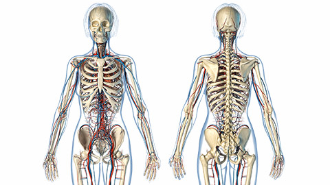
Kia ora and welcome to Patient Care 2. As you move through this topic, you will discover the seamless connection between Patient Care 2 and Anatomy and Physiology. These two topics complement each other and serve as building blocks for developing your understanding of:
- Anatomy (specifically looking at the body organ systems in the image above)
- Health disorders
- Pharmacology of medications used in treating health conditions
- Counselling patients on the use of medicines to treat disorders of different body systems.
Introduction
As a pharmacy technician, it is vital that you have a solid understanding of the human body, how it works and the disorders that can affect it. It's through this understanding that you'll be able to assist in the identification and management of various medical conditions. Combined with a clear understanding of pharmacology, you will be able to dispense, under supervision, medications that are appropriate, safe and effective in treating health disorders.
The knowledge and insights gained from this topic will empower you to communicate with your patients effectively, ensuring they comprehend the purpose, dosage, and potential side effects of their medications. All these actions, behaviours, skills and knowledge will ensure that you are able to provide an excellent level of pharmacy technician services to your patients, contributing to their recovery and well-being.
Me ruku ki te kaupapa, let’s dive into our topic!
Structural organisation of the body
Let’s explore how all the parts of the body are organised so that they work together and form the expected human anatomy. To do this, we will need to start small, with the extremely tiny particle called the atom. You may be familiar with what an atom is. Take a moment now to check in on your existing knowledge or research a definition.
Once you’ve done that, click the (+) symbol to reveal one definition of an atom and compare it to yours. Note: you click on the highlighted words in the definition to discover more.
So, how do we get from a single atom to a complete human body? Take a look at this graphic to get an overview.
Organ systems
The human body is grouped into organ systems, sometimes called body systems. You will see that many medical texts divide the body into 11 or 12 organ systems, but designating organs to organ systems can be complicated. This is because organs that “belong” to one system can also have important functions in another system. In fact, most organs contribute to more than one system.
For our purposes of relating the anatomy of body systems to the treatment of health disorders, we will look at the human anatomy and body systems in the following way:
- Cardiovascular system
- Nervous system
- Digestive system
- Endocrine system
- Respiratory system
- Reproductive and urinary systems
- Musculoskeletal and integumentary systems
- Eye, ear, nose, and oropharynx
- Immune system.
This week, we will investigate the cardiovascular system.
Cardiovascular system
Welcome to your learning on the cardiovascular (CV) system! To start, we'll do a quick activity to check your general knowledge of the CV system. Don’t worry if this information is new to you. Give it your best guess and take note of the correct answers.
The role of these structures
In very simple terms, the role of each structure is as follows:
- The heart: It pumps blood through blood vessels, ensuring that all cells receive the necessary oxygen and nutrients for their functions.
- Blood: The fluid that carries oxygen, nutrients and waste products.
- Blood vessels: The structures that contain and carry blood around the body.
Now, let’s take a closer look at the anatomy of each of these structures.
Anatomy of the heart
Where is the heart located in the body? It may not be exactly where you think it is!
| Many people think the heart is located on the left side of the chest, when in fact, it is in the middle of the chest cavity with the bottom, rounded end (the apex) tipped slightly towards the front of the body and towards the left-hand side. | 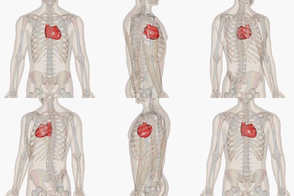 BodyParts3D/Anatomography, CC BY-SA 2.1 JP |
The heart is made up of specialised muscle tissue called cardiac muscle or myocardium. It consists of four chambers: two upper chambers called atria, which receive blood, and two lower chambers known as ventricles, responsible for pumping blood.
The heart also has four valves to ensure one-way blood flow: the tricuspid and mitral valves separate the atria from the ventricles, and the aortic and pulmonary valves regulate blood flow into major arteries. The heart's rhythmic contractions maintain blood circulation, supplying the body's tissues with oxygen and nutrients while removing waste products.
Watch: Heart Anatomy in 3 Minutes | Memorize parts of the heart (3:34 minutes)
Watch this video that describes the anatomy of the heart and the direction of the blood flow through the heart. Āta whakarongo, listen carefully to the names of the atria, ventricles, valves and vessels, and then test your recall in the activity following the video.
How well do you know the heart? Test your knowledge and drag the names of the parts of the heart to the correct locations.
Anatomy of blood
The next structure of the CV system is blood.
Watch: Blood, Part 1 - True Blood: Crash Course Anatomy & Physiology #29 (9:59 minutes)
Mātakitaki mai, watch this video to learn about the basic components of blood, blood donation, hemostasis, how bleeding works, antigens and blood types. Feel free to pause and take notes as you watch to help you retain any information that is new to you. You will also be given the opportunity to check your knowledge with a short quiz following the video.
Answer the following five questions to help summarise the blood crash course you’ve just experienced.
Oxygenated and deoxygenated blood
When we talk about blood, we refer to oxygenated blood and deoxygenated blood, each with its unique role:
- Oxygenated blood:
This is blood rich in oxygen, typically found in arteries. It is bright red and carries oxygen from the lungs to the body's tissues, providing them with the vital oxygen needed for energy production and body functions. - Deoxygenated blood:
Deoxygenated blood is low in oxygen and appears darker, often seen in veins. It returns from the body's tissues to the heart, where it is pumped to the lungs. There, it picks up oxygen and releases carbon dioxide, getting ready to become oxygenated again in a continuous cycle.
As you can see, the cardiovascular system relies on the cooperation of another body system, namely the respiratory system, to fulfil its vital function.
Anatomy of blood vessels
Blood vessels circulate blood around the body. The network of blood vessels is referred to as the blood circulatory system. It is a closed, continuous system. The body has another circulatory system, which is an open system that carries a fluid called ‘lymph fluid’. You will learn about this system later in your programme.
There are five main types of blood vessels in the blood circulatory system.
These are:
- Arteries
- Arterioles
- Capillaries
- Venules
- Veins
Journal post
Blood vessels
As you continue into Level 5 study, you will notice an increase in self-led learning activities as we ask you to conduct your own research to seek information. This activity will require you to put on your investigation hat to find out more about these five main blood vessel types.
- Complete the following Documentation tool activity.
- Download and save the completed activity.
- Create a new journal post titled ‘Week 26 - Blood vessels’.
- Upload the documentation tool to your journal post and publish it to ‘All course users’. This will allow your tutor to review your work and provide you with feedback.
Blood flow
Watch: The Pathway of Blood Flow Through the Heart, Animation (2:09 minutes)
Now that you have a clear idea of the anatomy of the CV system, head to YouTube to view this video from Alila Medical Media to see the blood flow as it travels through the heart. The Pathway of Blood Flow Through the Heart.
From this video, you will have noted that blood is pumped through both the left and right sides of the heart at the same time.
It is important to note that each time blood is pumped out of the heart to supply the body, a portion travels via the coronary blood vessels to the outside of the heart to feed the heart muscle.
All muscles need blood to function, and the heart muscle is no different. When the coronary vessels do not function as they should, it results in damage to the heart muscle. Like any damaged muscle, its function is then impaired. This can lead to serious and life-threatening health conditions.
We will talk more about these health conditions in Anatomy and Physiology.
In summary, the anatomical structures of the cardiovascular system work together to pump and circulate blood throughout the body. These actions ensure the delivery of oxygen function and the health of the entire body.
Self-directed learning activities
This week, it’s up to you to set yourself up for success! Complete the following activities to start Level 5 on a strong note.
activity 1 – Note-taking and organisation
Good note-taking can enhance your learning and save you time. Use the opportunity now to decide how you are going to create and save your own notes and learning activities for future reference. You may want to continue with whatever methods you used in Level 4 or take the opportunity to improve your methods. There are lots of ways to make your own notes, including Microsoft Word, Microsoft OneNote, Apple Notes, or even a good old-fashioned physical notebook. If you're after some inspiration for a new method, check out this article from Zapier: The 6 best note-taking apps in 2024.
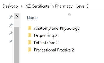
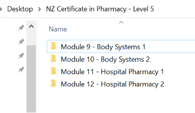
Think about where you save your documents, too. For example, you may want to create a folder on your device for each module, each topic, each week, or even each activity. Everyone’s brains work differently, so take a moment to think about the system that works best for you.
Here are some examples below:
By topic:
By module:
activity 2 – anatomical and directional terminology
It's time to add some more medical terminology to your ever-expanding healthcare vocabulary! Watch this video to hear how locations on the body are talked about.
In pharmacy, you may not use these words often, but it helps to have a bit of an awareness of directional terms when collaborating with other health professionals who may use them.
Some common terms you are likely to hear are anterior, posterior, superior and inferior.
Āta whakarongo, listen carefully to the video for these terms and note down any other terms that are new to you.
Watch: Anatomical Positional And Directional Terms (3:15 minutes)
Note: You will encounter a lot of new terminology throughout the rest of the course, so we suggest starting your own glossary of these terms!
That’s the end of Week 26 for this topic. Whakamihi, well done. You’ve covered a lot of content!
Nau mai, welcome to Anatomy and Physiology. You will notice that this topic and the topic of Patient Care 2 are inter-related. The learning from one topic will support the learning in the other.
Introduction
Our focus on this topic is on the application of an advanced level of human anatomy, physiology and medicines used in the treatment of health disorders. We have structured the learning in this topic into the following body systems in this order.
- Cardiovascular system
- Nervous system
- Digestive system
- Endocrine system
- Respiratory system
- Reproductive and urinary systems
- Musculoskeletal and integumentary systems
- Eye, ear, nose, and oropharynx
- Immune system and malignant disease.
The learning from this topic is crucial in preparing you for your role as a pharmacy technician. You will gain advanced knowledge of health disorders that affect each body system and the medications used in their treatment. This knowledge is invaluable, enabling you to ensure the safe and effective use of medicines. You'll be equipped to feel confident in making informed decisions when dispensing drugs, educating patients about their medications, and identifying potential interactions or contraindications.
Journal post
What are anatomy and physiology?
Let’s begin by defining anatomy and physiology.
- Create a new journal post titled ‘Week 26 - What are anatomy and physiology?’
- Write your own definitions for anatomy and physiology. If you need a prompt to get started, think about how the words ‘structure’ and ‘function’ might relate to your definitions.
- Publish your journal post to ‘All course users’ so that your classmates and tutor can see your definitions. You might look to check out your classmates' posts too!
- Save the permalink to your Index of Journal Posts.
Homeostasis
For the human body to function as expected, all the structures of the body must be present and organised in the expected way. The body must also work together in a way that keeps it in the functioning condition called homeostasis (this is different to hemostasis!).
What does homeostasis mean? Check your understanding (or have a guess!).
Therefore, we would say homeostasis means keeping the body in the same or similar state at all times so that it can function correctly despite the changes the body encounters from the environment. For example, the human body has a temperature set point as we know. This set point is 36.5oC – 37.5oC. This is the optimal temperature range for our bodies to function.
When the external environment, like air temperature, changes from this set point, our body uses physiological compensatory mechanisms to return its internal temperature to its set point to achieve homeostasis.
Using the example of air temperature changes, let’s look at the compensatory mechanisms that return the body to a homeostatic state.
Scenario
Air temperature
Read this scenario and then consider the following questions.
Allen is outside in his garden. It is a hot summer’s day, and he is mowing the lawn. The weather report says the temperature is currently 28oC. Allen’s body temperature has risen, and he feels hot and uncomfortable.
- How will Allen’s body compensate for this rise in body temperature?
- What will the body do to return its temperature to its set point?
How did you go? Did you know the information already, or did you need to conduct some research? Check your answers by clicking on the (+) symbol below.
- The blood vessels in Allen’s body will dilate (open) to allow excess heat from inside the body to escape through the skin. His body will also produce sweat.
- When sweat evaporates from the surface of the body, it produces a cooling effect, which contributes to lowering the body temperature.
Compensatory mechanisms
There are many compensatory mechanisms that the body uses to maintain its many functions within specific set point ranges.
These body functions include:
- Temperature
- Blood pressure
- Heart rate
- Respiratory rate
- Blood glucose levels
- Oxygen levels
- Electrolyte balance
- Fluid balance
- pH balance
- Waste elimination.
Cardiovascular anatomy and physiology
Let’s take a moment now to review the cardiovascular (CV) anatomy that we looked at this week in Patient Care 2.
Journal post
This activity allows you to check your knowledge of the anatomy of the heart, the circulatory system and coronary circulation.
- Download and complete the worksheet: Week 26 - AP - Cardiovascular Anatomy - Worksheet. (Note: it is a ZIP file, so you will need to open it to find the worksheet.)
- Save the completed worksheet and upload it to your journal under the title: ‘Week 26 – CV Anatomy’.
- Publish to ‘All course users.’ This will allow your tutor to check your work and ensure that you're on track!
- Save the permalink to your Index of Journal Posts.
- Make sure to check back on your post later to see if your tutor has left any feedback for you.
As you are aware, the cardiovascular system depends on the respiratory system for oxygenating blood and removing carbon dioxide. As well as the lungs playing a crucial role in the CV system, so too do the kidneys. The kidneys help to regulate blood pressure and maintain the body’s fluid balance. As you progress through your learning, you'll explore the structure, function, and interconnections of the kidneys with the CV system as well as other body systems.
Circulatory pathways
There are three circulatory pathways in the CV system.
Watch: Systemic and Pulmonary Circulation (29 seconds)
Watch this video that shows these three blood pathways, and then complete the following activity to confirm your understanding.
The cardiac cycle
The cardiac cycle is the sequence of events that occur in the heart from the beginning of one heartbeat to the beginning of the next heartbeat. During this time, both atria and ventricles contract and then relax. As we know from our previous knowledge, the average number of heartbeats for an adult is 70-80 beats per minute.
The cardiac cycle is essential for pumping blood through the circulatory system, ensuring the delivery of oxygen and nutrients to the body's tissues and the removal of waste products.
Cardiac conduction
The heart is made up of cardiac muscle tissue, which is different from the other muscle tissue found in the body. The cells that form cardiac muscle tissue are interconnected, which allows them to transmit electrical signals that coordinate the heart's rhythmic contractions.
These electrical signals are triggered by the heart's natural pacemaker called the sinoatrial (SA) node. The electrical signals then travel through the heart in a specialised pathway called the cardiac conduction system.
Watch: Electrical Conduction System of the Heart (3:01 minutes)
This video illustrates the cardiac conduction system in action. As you watch, notice the opening and closing of the heart valves in the chambers and the large vessels at different times during the cardiac cycle. After you watch the video, complete the sequencing activity to support your learning.
Tip
You may want to watch this video more than once in order to see all that is happening with each heartbeat.
Blood pressure and the cardiac cycle
What is blood pressure, and how does it relate to the cardiac cycle?
Take a moment to consider this and find out what the terms ‘systole’ and ‘diastole’ mean. Once you have written your own definitions in your notes, click on the (+) symbol for an explanation.
Blood pressure is a measurement of force. It is the force of blood pushing against the walls of the arteries as the heart pumps it around the body. A blood pressure reading is made up of two numbers. Each number is a measurement of force at different phases of the cardiac cycle. This force is measured in millimetres of mercury or mmHg.
The two numbers are as follows:
- Systole: This is the period of time in the cardiac cycle where the ventricles contract and pump blood out of the heart into the large vessels on its way to the rest of the body. The force used by the ventricles to pump blood with each heartbeat causes a rise in pressure inside the arteries of the body. This pressure is called ‘systolic pressure’. It is measured and recorded as the first or top number in a blood pressure reading.
- Diastole: This is the period of time in the cardiac cycle where the ventricles relax and refill between contractions. During this time between heartbeats, the pressure on the walls of the arteries drops. This pressure is called the ‘diastolic pressure’. Diastolic pressure is the second or bottom number in a blood pressure reading.
Blood pressure
To help you to retain this information, answer the following two multichoice questions. The quiz will automatically advance once you select your answer.
The answers to the above questions are guides only. Guidelines for the categorisation of ‘normal’ blood pressure and the cut-off points for different degrees or severity of high and low blood pressure may differ from country to country. A health practitioner will also look at many other factors in assessing a patient's blood pressure.
Journal post
Blood pressure categorisations
What do you think these factors might be?
- Create a new journal post titled: ‘Week 26 - Blood pressure categorisations’.
- Write your answer to this question in your journal post.
- Publish to ‘All course users’ so your tutor can read your work and leave feedback.
- You'll be in the rhythm of this now, but remember to save the permalink to your Index of Journal Posts! You'll thank your past-self for this later.
As we have seen, both the anatomy (structures) and the physiology (function) of the CV system are responsible for maintaining homeostasis and sustaining life. The anatomical components work together with the physiological processes to regulate blood flow, ensure the delivery of oxygen and nutrients to tissues, and facilitate the removal of waste products.
Having explored the cardiovascular system's anatomy and physiology, let's delve into the disorders impacting this body system and the medications used to treat the disorders.
CV disorders and treatments
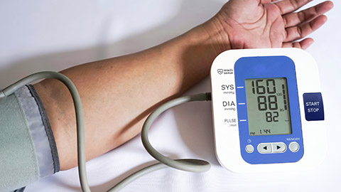
Hypertension (Mate pēhanga toto)
High blood pressure is known as hypertension. Hypertension affects 1 in 5 people in this country and can lead to a wide range of CV disorders (Healthify 2022).
Watch: Hypertension - causes, symptoms, diagnosis, treatment, pathology (6:16 minutes)
This video is helpful in understanding the factors that affect blood pressure and some of the causes, risk factors and signs and symptoms of hypertension. Once you’ve watched the video, complete the following journal activity to reflect on your learning.
Note: The parameters for blood pressure readings and classifications may differ between countries.
Journal post
Hypertension
- Complete the following Documentation tool activity.
- Download and save the completed activity.
- Create a new journal post titled ‘Week 26 – Hypertension’.
- Upload the documentation tool to your journal post and publish it to ‘All course users’. This will allow your tutor to review your work and provide you with feedback.
Treatments
The last part of the video touched on lifestyle changes and medication as treatments for hypertension.
| Lifestyle changes | Medication |
|---|---|
|
Lifestyle changes play a role as preventive measures and in contributing to the ongoing treatment of hypertension. Changes include the following:
|
Several therapeutic groups of medicines exist for treating hypertension. These include:
According to bpac NZ (2023), in New Zealand, the first line of medicinal treatments for hypertension are:
Beta-blockers are no longer used as first-line treatment unless indicated for a specific clinical reason. |
Self-directed learning activity
In this self-directed activity, you will learn more about the first line of antihypertensives (medicines used to lower high blood pressure) and will look at Angiotensin-Converting-Enzyme (ACE) inhibitors.
- Create a new journal post titled: ‘ACE Inhibitors.’
- Record your answers to the following two activities in the journal post.
- Save the post and publish it to ‘All course users’ so your tutor can check out your mahi.
- Save the permalink to your Index of Journal Posts.
activity 1
Briefly explain how ACE inhibitors work to reduce high blood pressure. Use the following video and resources to assist you.
Watch: How do ACE inhibitors work? (2:11 minutes)
Watch this video for a simple and clear explanation.
Other resources
You may also like to search these websites for relevant information.
Activity 2
Enalapril is an example of an ACE inhibitor used to lower blood pressure. Using the resources available to you, find the answers to the following questions about this medicine:
- When prescribed for the treatment of hypertension, what is the typical dose and frequency of dosing for enalapril?
- What are common side effects?
- How can these side effects be managed?
Resources
That brings us to the end of Week 26 for Anatomy and Physiology. Mālō lava! Well done! We hope you have enjoyed your learning this week and can see the close connection between this topic and Patient Care 2.
