Just like the rest of the body, bones, circulation, muscles and nerves enable the body to function as it should.
Arches of the foot
The arches of the foot are created by the formation of the bones and joints and supported by ligaments.
These arches support the weight of the body and help to preserve balance when we walk on uneven surfaces.
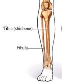
The lower leg is made up of 2 long bones, the tibia and the fibula. These bones have joints with the upper leg (at the knee) and with the foot (at the ankle).
Having 2 bones in the lower leg– as with the forearm– allows a greater range of movement to be achieved at the ankle.
The bones of each lower limb are also formed from 30 bones and include
- Femur
- Patella
- Tibia
- Fibula
- Tarsals
- Metatarsals
- Phalanges
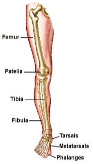
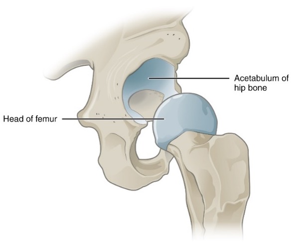
The femur, also known as the thigh bone, is the longest and strongest bone in the body and forms our thigh. It articulates with the pelvic bones at the acetabulum and extends down towards the knee joint where it articulates with the bones of the lower leg. The femur slants medially as it runs downward to join with the leg bones, this brings the knees in line with the body’s centre of gravity.
The patella is commonly known as the kneecap. The area behind the patella is called the popliteal. It is a thick movable anterior bone that protects the knee area, between the femur and tibia. Strong cartilage allows it to be moveable. It articulates with the femur to form the knee joint.
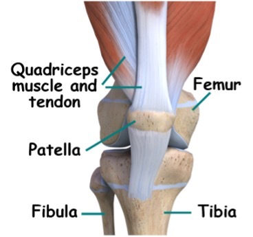
These are the weight bearing bones of the body and form the lower leg. The tibia, commonly known as the shinbone, is the larger or thicker bone and is on the big toe side of the lower leg, or medial to the fibula.
The fibula is the fine sticklike bone that is on the little toe side of the lower leg, or lateral to the tibia.
An easy way to remember them
TIBIA – Thick
FIBULA – Fine
The bones fit together to form arches which help to support the foot and to absorb the impact when we walk, run and jump.
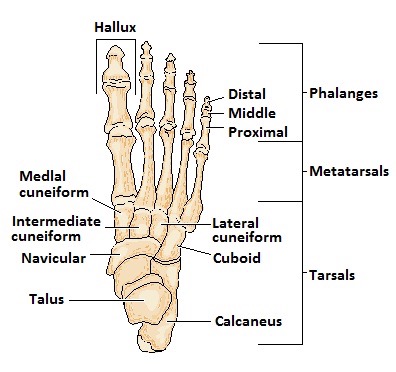
The foot supports our body weight and acts as a lever to propel the body forwards during walking and is made up of 26 bones.
- 7 tarsals, ankle bones. The calcaneus is commonly known as the heel and the talus is known as the anklebone.
- 5 metatarsals, bones in the front of the foot.
- 14 phalanges, toe bones, the big toe is often referred to as the hallux.
Muscles of the Foot and Lower Leg
The muscles of the foot work together, to help move the body when walking and running. In a similar way to the movement of the hand, primarily muscles in the lower leg move the foot. These pull on tendons, which in turn move the feet and toes.
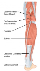
Our leg muscles enable us to run, jump, hop and walk. Like arm muscles there are both muscles, which are individual, or grouped together because of their action and connections. The main muscles in the legs are
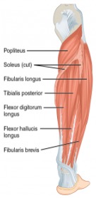
Sits at the posterior lower leg, known as the calf muscle, flexes the knee, plantar flexes the foot and flexes the leg.
Lies under the gastrocnemius and plantar flexes the foot.
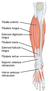
Runs alongside the tibia bone. Inverts (turns inwards) and dorsiflexes the foot.
As per arm muscles, there are a number of these in the legs, they either
- Extend or lengthen a muscle
- Flex or shorten or contract a muscle
Tendons and Muscles in the Foot and Ankle
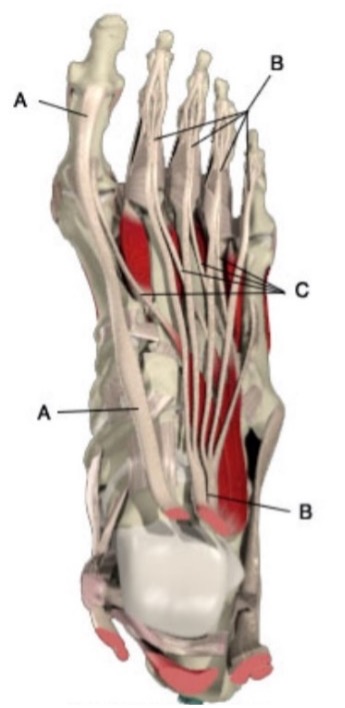
In the foot diagram to the right, the main tendons on the top of the foot are
A. Extensor hallucis longus: dorsi flexes the big toe and dorsiflexes the foot (pulls up the big toe and the foot).
B. Extensor digitorum longus: dorsi flexes the small toes and dorsiflexes the foot (pulls the little toes up and pulls the foot up).
C. Extensor digitorum brevis: dorsi flexes the small toes (pulls the small toes up).
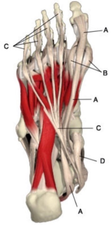
In the foot diagram to the right, the main tendons on the bottom of the foot are
A. Flexor hallucis longus: plantar flexes the hallux and the foot (brings the big toe and flexes the foot down).
B. Flexor hallucis brevis: plantar flexes the hallux (brings the big toe down).
C. Flexor digitorum longus: plantar flexes the toes and the foot (brings the little toes down and flexes the foot down).
D. Posterior tibialis: inverts and plantar flexes the foot (brings the foot in and down).
Our circulatory and lymphatic system has the same functions, no matter where it is place in our body.
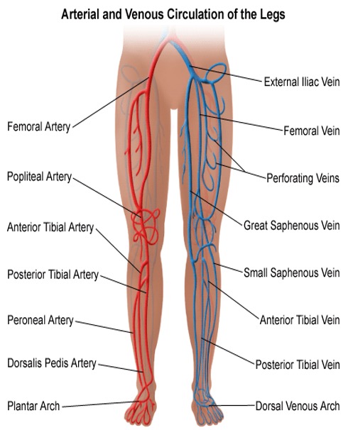
The femoral artery is the main blood supply to the legs; from this the popliteal artery branches off at the knee and then divides into the anterior and posterior tibial arteries which supply the leg and foot. The anterior tibial artery finishes with the dorsalis pedis artery which supplies the foot.
The important veins are located almost parallel with the arteries and take the same names as the arteries. While the arteries are found deep in the tissues, the veins lie nearer to the surface of the arms and hands.
The main arteries and veins of the lower legs are
- Femoral Artery
- Anterior tibial Artery
- Posterior tibial Artery
- Saphenous Vein
- Femoral Vein
This runs from the groin to the back of the knee where it becomes the popliteal artery.
This starts at the popliteal artery and travels down the front of the tibia and fibula, at the ankle it divides again into smaller arteries that supply the foot.
Runs from the popliteal artery down the back of the leg to the ankle, where it becomes the plantar artery.
There are two of these veins, great and small. The great saphenous is the longest vein in our body, running from the top of our foot, along the inside of our leg to the groin, where it joins into the femoral vein. The small saphenous vein runs from the back of the ankle to behind the knee, where it joins the popliteal vein.
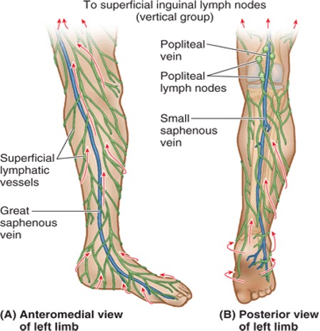
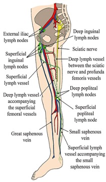
Like our arms, lymphatic vessels are deep and superficial, and have the same or similar names as the veins they work closely with. There are many smaller lymphatic capillaries that drain into vessels, to be filtered, before eventually returning to venous blood circulation. The main lymphatic nodes in our legs are
- Popliteal nodes
- Inguinal nodes
There are between 5 – 7 nodes that sit behind the knee joint, in the popliteal area. They filter all lymph from the feet and lower legs. When your legs feel heavy and tired, massaging, or sitting with your feet elevated can help with drainage, alternatively brisk movements; fast walking, jogging on the spot etc, can stimulate the circulation, in turn stimulating the lymph movement. Massaging tired legs is a great way to alleviate this. The area behind and around the knee can be tender, always check with your client, make sure the pressure is ok.
There are a few sub-divisions of these nodes, but they all sit in the general region of the groin.
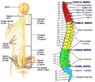
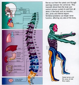
Our body has nerve endings everywhere. These are the receptors that either send or receive messages in response to internal and external stimuli. Nerves are our internal communication system that keeps us safe and functioning. When it is cold, nerve receptors in the skin send messages to the brain, which is interpreted, and another message is sent back to initiate a response, e.g. shivering, the quick contraction and relaxation of the muscles stops heat from escaping our body, erector pili muscle contracts, making hairs stand on end trapping heat against the surface of the skin.
We have millions of nerves pathways in our body. Nerves run through the spinal cord, then branch out to specific areas in our body.